MRI vs fMRI: What Is the Difference?
A standard MRI shows what the brain looks like. A functional MRI shows which parts of it are working during a task. They use the same kind of scanner but answer very different questions.
If you have read about brain scans, you may have come across both MRI and fMRI and wondered whether they are the same thing. They are closely related. Both are carried out on an MRI scanner, and both involve lying still inside it while pictures are taken. The difference is in what the pictures show and why they are taken.
This guide explains the difference, where functional MRI is used, what happens during one, and why it is a specialist hospital test rather than a routine scan you would arrange privately.
Standard MRI: a picture of structure
The NHS describes an MRI scan as a scan that takes detailed pictures of the inside of your body. An MRI scanner uses a strong static magnetic field, gradient magnetic fields that switch rapidly, and radio waves, the three fields that MHRA safety guidance sets out for MRI equipment. No ionising radiation is involved.
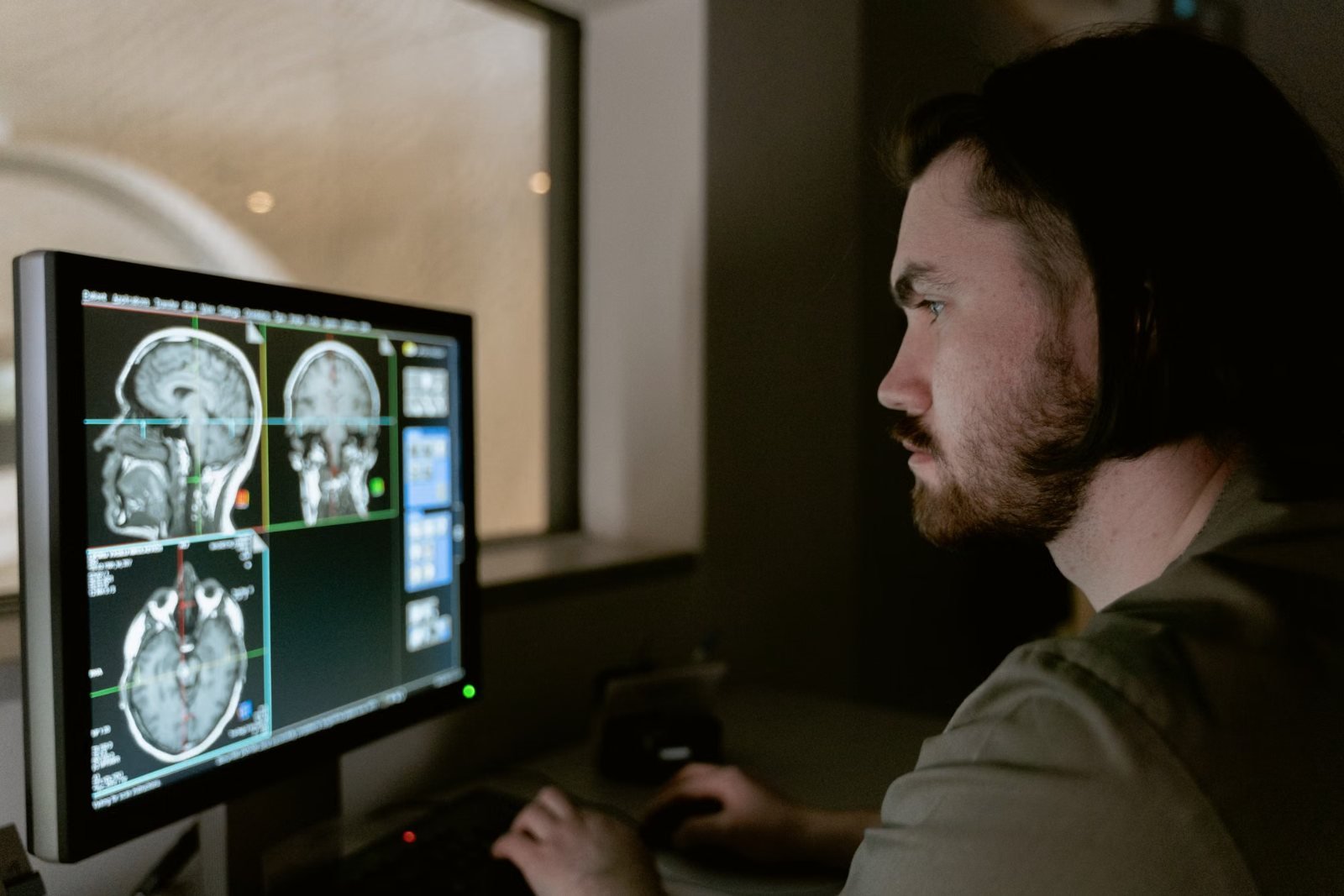
A standard brain MRI shows the structure of the brain: its shape, the tissues and the fluid spaces around it. Doctors use it to look for changes such as damage, inflammation or a growth. Our guide to what a brain MRI shows explains more. A structural scan does not show how well the brain is working, and it is not a memory or thinking test.
Functional MRI: a picture of activity
Great Ormond Street Hospital explains that while a regular MRI shows structure, a functional MRI scan shows which parts of the brain are activated when certain tasks are carried out, such as language, memory and movement. It does this using a technique called blood oxygenation level dependent (BOLD) imaging, which picks up changes in blood flow that go with brain activity.
In other words, fMRI tracks a change in the blood supply that goes with activity, rather than the activity itself, and builds a map of the areas that responded while you did a task.
Where fMRI is used: surgery planning
A consensus paper on clinical fMRI, published in Aperture Neuro in 2025, says the main clinical applications of fMRI for individual patients include assessing risk before an operation and mapping critical areas of the brain for surgical planning.
In practice, that usually means planning brain surgery, for example for epilepsy. University College London Hospitals describes fMRI as identifying the parts of the brain involved in functions such as speech, reading, memory, vision, sensation and limb movement, so that surgeons know which areas to protect. Great Ormond Street Hospital says it can be extremely useful when planning brain surgery for the same reason.
The same consensus paper is candid that fMRI's usefulness for clinical language mapping is still debated, partly because centres differ in how they acquire, analyse and interpret the data. That is one reason it is carried out by specialist teams.
Where fMRI is used: research
fMRI is also a research tool. NHS research summaries published by the Health Research Authority include, for example, a study using fMRI to examine how memory networks adapt after epilepsy surgery.
What happens during an fMRI
The safety checks are the same as for any MRI, because the same scanner is used. The difference is that you are asked to do things during the scan. Great Ormond Street Hospital mentions tasks such as answering questions or tapping a finger, and UCLH describes thinking of words beginning with a particular letter, or moving a hand, while looking at pictures and words.
Great Ormond Street Hospital tells families its fMRI scans can take up to two hours, and that the scanner makes a continuous, quite loud knocking sound, so ear protection is provided. It describes the scan as painless with no lasting effects. A standard MRI scan, by comparison, usually takes between 15 minutes and 1 hour, according to the NHS. Our guide to what to expect during an MRI scan covers the general experience.
Can you have an fMRI privately?
fMRI is a specialist test arranged by a hospital team, usually as part of planning surgery, rather than a routine scan. If you are thinking about a private brain scan, check with the clinic exactly what a listed scan includes. A standard brain MRI shows structure, not function.
MRI does not use ionising radiation, so IR(ME)R does not apply. Many private clinics accept self-referral, usually after their own safety screening, but each clinic sets its own policy. Each scan is reported by an appropriately qualified clinician, and the clinic will tell you how long its report takes. It is worth talking to your GP first about whether a scan is needed; see our guide to seeing a GP before a scan, the brain MRI scan page, or compare MRI clinics near you.
Call 999 if you have a headache that came on suddenly and is extremely painful, or weakness or difficulty moving one side of your body or face. These are emergencies, not reasons to wait for a scan.
Common questions
Is fMRI done on the same machine as an MRI?
Yes. fMRI is carried out on an MRI scanner. The difference is in the way the images are taken and analysed, using the BOLD technique to track changes linked to blood flow while you do tasks.
What does fMRI show that MRI does not?
A standard MRI shows the structure of the brain. An fMRI shows which areas become active during tasks such as speaking, remembering or moving, which helps surgeons plan operations near important areas.
Can I ask for an fMRI instead of a normal brain MRI?
fMRI is a specialist test used mainly to plan brain surgery and in research. It is arranged by a specialist team when it is needed. If you are worried about your brain health, start with your GP.
Is fMRI safe?
It uses the same scanner as a standard MRI, so the same safety screening applies, and there is no ionising radiation. Great Ormond Street Hospital describes it as painless with no lasting effects. The scanner is loud, so ear protection is provided.
How long does an fMRI take?
It depends on the tasks involved. Great Ormond Street Hospital tells families its fMRI scans can take up to two hours. A standard MRI usually takes between 15 minutes and 1 hour, according to the NHS.
References
- Functional magnetic resonance imaging (fMRI). Great Ormond Street Hospital (NHS). Accessed 6 October 2026.
- A guide for patients considering epilepsy surgery. University College London Hospitals (NHS). Accessed 6 October 2026.
- Consensus recommendations for clinical functional MRI applied to language mapping. DOI 10.52294/001c.128149. Aperture Neuro (Voets et al., 2025). Accessed 6 October 2026.
- Cognitive fMRI in Temporal Lobe Epilepsy (research summary). Health Research Authority (NHS). Accessed 6 October 2026.
- MRI scan. NHS. Accessed 6 October 2026.
- Magnetic resonance imaging equipment in clinical use: safety guidelines. MHRA (gov.uk). Accessed 6 October 2026.
- Migraine. NHS. Accessed 6 October 2026.
This article is general information, not medical advice, and it does not replace an assessment by a clinician. If you have symptoms that worry you, speak to your GP or call 111. In an emergency, call 999.