Breast MRI scan vs mammogram: what each test does and who needs which
A mammogram uses X-rays, while a breast MRI uses a magnet and usually a contrast injection. Here is what each test does, who breast MRI is for, and why mammography is still the screening test in the UK.
Written by the FindScan Editorial Team and reviewed by our Medical Advisor, Dr Mohamed Najjar
Reviewed
Mammograms and breast MRI scans both create pictures of the inside of the breast, but they work in very different ways and are used for different people. This guide explains what each test does, who a breast MRI scan is usually for, and why mammography is still the main screening test in the UK. If you are comparing private options, see our private mammogram page and our private MRI scan page.
The short version
- Mammogram: an X-ray of the breast. It is the test used in the NHS Breast Screening Programme.
- Breast MRI: uses a strong magnet and radio waves, with no X-rays. It is usually saved for specific situations, such as people at very high risk, very dense breasts, possible implant problems, or planning treatment after a cancer diagnosis.
Neither test is simply better than the other. Each has strengths and limits, and the right choice depends on your age, your risk and the reason for the scan.
What a mammogram does
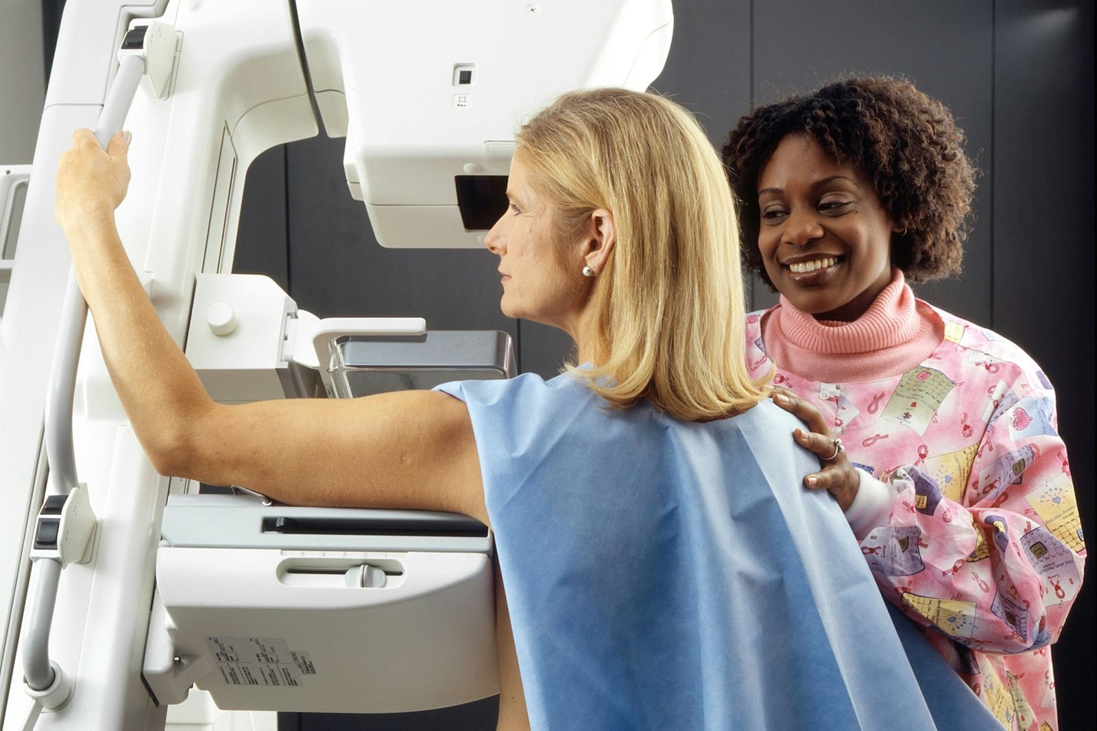
A mammogram uses X-rays to take pictures of the breast. In NHS screening, you'll have 2 mammograms taken for each breast. Each image takes a few seconds, and the whole appointment usually takes no longer than 30 minutes.
The NHS offers screening because it can save lives from breast cancer. Finding cancer early may mean simpler and more effective treatment.
Mammograms do have limits. The GOV.UK screening leaflet says that screening does not always find a cancer that is there. It also explains that the main risk of screening is that it may find a cancer that would never have caused harm, and that mammograms involve a small amount of radiation.
Breast density matters too. Breast Cancer Now explains that younger women are not usually offered mammograms because they are more likely to have dense breast tissue, which can make mammogram images less clear.
What a breast MRI does
MRI uses a magnetic field and radio waves rather than X-rays. Breast Cancer Now notes that MRI doesn't expose the body to x-ray radiation.
A breast MRI is a longer and more involved test than a mammogram. The breast unit at Bedfordshire Hospitals NHS Foundation Trust explains that it usually involves:
- an injection of a contrast medium called gadolinium into a vein
- lying on your front on a moving table that slides into the scanner
- a scan time of around 45 to 60 minutes
- quite a lot of noise, so you will be given headphones
If you are nervous about MRI, our guides on what to expect during an MRI scan and MRI with contrast may help.
Who breast MRI is used for
People at very high risk of breast cancer
Some people have a much higher than average chance of developing breast cancer, often because of an inherited gene change. NICE guidance on familial breast cancer (CG164) recommends annual MRI surveillance for:
- women aged 30 to 49 with a known BRCA1 or BRCA2 gene change, or a greater than 30% chance of carrying one
- women aged 20 to 49 with a known TP53 gene change, or a greater than 30% chance of carrying one
The NHS Breast Screening Programme runs a very high risk service. Its protocols include people with certain gene changes and some who had radiotherapy to breast tissue for Hodgkin or non-Hodgkin lymphoma at a young age. In this service, women have both MRI and mammography up to and including the age of 50. After that, women aged 50 or over have mammography with or without MRI, depending on a review of their breast density.
Breast Cancer Now explains that eligibility for this kind of screening is usually decided by a genetics team. If you are worried about your family history, your GP is the place to start.
Dense breasts
Dense breast tissue can make a mammogram harder to read. NICE says clinicians should explain the possibility that mammography might miss a cancer in women with dense breasts. For women aged 50 and over without a TP53 gene change, NICE says MRI surveillance should not be offered unless mammography has shown a dense breast pattern.
The Bedfordshire breast unit also lists very dense breast tissue as a reason for MRI, especially when mammograms may not give much information.
Breast implants
MRI can help when there is a question about an implant. The Bedfordshire breast unit says MRI may be used if an ultrasound has suggested that there may be an implant leak.
Planning treatment after a diagnosis
After a diagnosis of breast cancer, MRI is not used for everyone. NICE guidance on early and locally advanced breast cancer (NG101) says do not routinely use MRI of the breast as part of the assessment before surgery. Instead, it recommends offering MRI to people with invasive breast cancer when:
- the extent of the cancer is not clear from examination, mammography and ultrasound
- breast density makes an accurate mammogram assessment difficult
- breast-conserving surgery is being considered for invasive lobular cancer, to check the size of the tumour
Bedfordshire Hospitals adds that MRI can be used to monitor how a cancer responds to treatment given before surgery.
The limitations of breast MRI
MRI is very sensitive, which is both a strength and a weakness. Bedfordshire Hospitals says breast MRI can often show up areas in the breast tissue which turn out to be harmless. These may need an ultrasound and a tissue sample to be certain.
A patient leaflet from Liverpool University Hospitals NHS Foundation Trust says about one in ten women who have a breast MRI are asked to come back for more tests. It reassures readers that most women who are called back will not have breast cancer.
Being called back can be worrying. NICE says people offered surveillance should be told about the possible psychological impact of a recall visit, along with other benefits and risks.
Other practical limits include the need for a contrast injection and the time spent lying still in the scanner. The NHS very high risk protocols note that if someone cannot have gadolinium or declines contrast, MRI should not be performed, and doctors will decide on another option such as mammography.
Why mammography remains the screening test
The UK National Screening Committee recommends breast screening. Its current recommendation is based on its last review in December 2019. The committee is looking at screening for women at higher risk, including risk due to dense breast tissue, but no changes to the programme have been made.
For most women, the NHS screening programme uses mammography. Breast MRI is added only for specific groups at very high risk, as set out in the NICE and NHS protocols above. Its tendency to pick up harmless changes, the need for contrast and the longer scan time are some of the reasons it is kept for people who are most likely to benefit.
Comparing the two at a glance
- How it works: mammogram uses X-rays; MRI uses a magnet and radio waves.
- Radiation: mammogram involves a small dose; MRI involves none.
- Contrast injection: not used for a standard mammogram; usually needed for breast MRI.
- Time: mammogram appointment up to about 30 minutes; MRI scan around 45 to 60 minutes.
- Main use: mammogram for screening and diagnosis; MRI for very high risk surveillance and specific problem solving.
If you have symptoms
If you notice a change in your breast, please speak to your GP as soon as possible, even if you have recently had screening. The GOV.UK leaflet gives this advice. Your GP can refer you to a breast clinic, where the right tests can be chosen for you.
This article is general information, not medical advice. Please speak to your GP or the clinician who referred you about any symptoms or results, and if your symptoms are urgent, call NHS 111 or 999.
Finding a private scan
You can compare clinics offering mammograms and MRI scans on FindScan, where each clinic lists its own price, and send an enquiry to the clinic you choose. The clinic will contact you to confirm details and the final price. A mammogram uses ionising radiation, so it must be justified and needs a referrer under IR(ME)R; many private clinics can arrange this through their own doctor. MRI does not use ionising radiation, and some clinics accept self-referral for MRI, but this depends on the clinic and the reason for the scan.
Common questions
Is a breast MRI better than a mammogram?
Not simply better. Breast MRI is very sensitive, but it often picks up areas that turn out to be harmless, which can lead to extra tests. It also usually needs a contrast injection and takes longer. Mammography is the test used in NHS breast screening, and MRI is kept for specific groups, such as people at very high risk.
Who should have a breast MRI?
NICE recommends annual MRI surveillance for some women at very high risk, such as those aged 30 to 49 with a BRCA1 or BRCA2 gene change, or aged 20 to 49 with a TP53 gene change. MRI may also be used for very dense breasts, a suspected implant leak, or planning treatment after a cancer diagnosis in specific situations.
Does a breast MRI use radiation?
No. Breast MRI uses a strong magnetic field and radio waves, so it does not expose you to X-ray radiation. A mammogram does use X-rays, although the dose is small. Breast MRI usually involves an injection of a contrast medium called gadolinium, and the scan typically takes around 45 to 60 minutes.
Why might I be called back after a breast MRI?
Because MRI is so sensitive, it often shows areas that need a closer look. A Liverpool University Hospitals leaflet says about one in ten women who have a breast MRI are asked back, and that most women who are called back will not have breast cancer. Further tests may include an ultrasound, extra mammogram views or a small tissue sample.
Can I have a breast MRI if I have dense breasts?
Dense breast tissue can make mammograms harder to read, and MRI is sometimes used for this reason. NICE says MRI surveillance should not be offered to women aged 50 and over without a TP53 gene change unless mammography has shown a dense breast pattern. The UK National Screening Committee is reviewing screening for dense breasts, but no changes have been made.
Do I need a referral for a breast MRI?
MRI does not use ionising radiation, so the IR(ME)R referral rules that apply to mammograms do not apply in the same way. Some private clinics accept self-referral for MRI, but this depends on the clinic and the reason for the scan. A mammogram always needs a referrer, and many private clinics arrange this through their own doctor.
References
- NHS breast screening: helping you decide. GOV.UK. Accessed 12 October 2026.
- Familial breast cancer (CG164): Recommendations. NICE. Accessed 12 October 2026.
- Early and locally advanced breast cancer: diagnosis and management (NG101): Recommendations. NICE. Accessed 12 October 2026.
- Protocols for surveillance of women at higher risk of developing breast cancer. GOV.UK. Accessed 12 October 2026.
- Breast MRI (Magnetic Resonance Imaging). Bedfordshire Hospitals NHS Foundation Trust. Accessed 12 October 2026.
- Attending breast assessment clinic following your MRI scan (PIF 1754). Liverpool University Hospitals NHS Foundation Trust. Accessed 12 October 2026.
- Family history of breast cancer: managing your risk. Breast Cancer Now. Accessed 12 October 2026.
- Breast cancer: screening recommendation. UK National Screening Committee (GOV.UK). Accessed 12 October 2026.
This article is general information, not medical advice, and it does not replace an assessment by a clinician. If you have symptoms that worry you, speak to your GP or call 111. In an emergency, call 999.