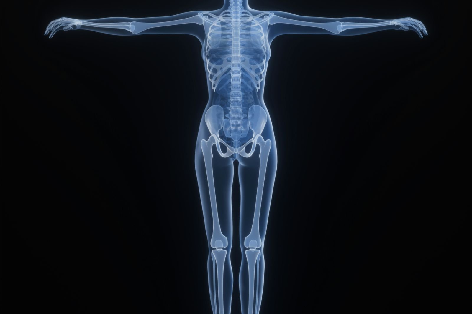
Will an X-ray show arthritis?
An X-ray can show some of the changes caused by arthritis, especially osteoarthritis in the knee and hip. But many people with typical symptoms are diagnosed without one, and an X-ray cannot tell you how much pain you will have.
Written by the FindScan Editorial Team and reviewed by our Medical Advisor, Dr Mohamed Najjar
Reviewed
The short answer
Yes, an X-ray can show some of the changes caused by arthritis. In osteoarthritis, Versus Arthritis explains that X-rays may show bony spurs called osteophytes, narrowing of the space between the bones and calcium deposits in the joint.
But an X-ray is not always needed. The NHS says that to diagnose osteoarthritis, a GP will first ask about your symptoms and examine your joints, and that further tests such as X-rays or blood tests are not usually necessary, although they may be used to rule out other causes. If your clinician does want imaging, a private X-ray is one option, but the test always needs a referral and must be justified, as explained below.
What an X-ray shows in osteoarthritis
Osteoarthritis affects the smooth cartilage that lines the ends of the bones and the bone around the joint. Versus Arthritis describes two main changes in the knee: the cartilage becomes rough and thin, and the bone at the edge of the joint grows outwards, forming bony spurs called osteophytes.
An X-ray shows bone clearly, so it can pick up the effects of these changes. A radiologist reading an X-ray for osteoarthritis may look for:
- Joint space narrowing: the gap between the bones looks smaller than expected. Because the cartilage becomes thinner in osteoarthritis, the bones can sit closer together.
- Osteophytes: bony spurs at the edges of the joint.
- Calcium deposits: small deposits of calcium within the joint.
Soft tissues such as cartilage, tendons and muscles do not show up in the same way. Versus Arthritis notes that an MRI scan will show the soft tissues and changes in the bone that cannot be seen on a standard X-ray, although MRI is rarely needed for osteoarthritis.
Knee X-ray and arthritis
The knee is one of the joints most often affected by osteoarthritis. Versus Arthritis lists the knees, hips, hands, spine and big toes as the most common sites, and notes that it often affects both knees, because the knee carries your body weight and copes with a lot of twisting and turning. A knee X-ray can show the bony changes described above, and the exact views taken depend on what your clinician asks for.
The radiation dose from a knee X-ray is very low. UK Health Security Agency (UKHSA) figures put X-rays of limbs and joints (except the hip) at less than 0.01 mSv, equal to less than 1.5 days of natural background radiation.
Hip X-ray and arthritis
A hip X-ray can show the same kinds of changes in the hip joint, such as narrowing of the joint space and bony spurs. The dose is higher than for a knee, but it is still low. UKHSA gives a typical dose for a hip X-ray of 0.3 mSv, equal to about 7 weeks of natural background radiation. For comparison, UKHSA says the average person in the UK receives about 2.1 mSv a year from natural background radiation.
Why X-ray results do not always match symptoms
One of the most important things to know about arthritis and X-rays is that the picture and the pain do not always line up. Versus Arthritis says X-rays are not a good indicator of how much pain or disability you are likely to have. Some people have a lot of pain from minor joint damage, while others have little pain from severe damage.
This is one reason why doctors focus on your symptoms, how the joint moves and how it affects your daily life, rather than relying on the X-ray alone.
Making sense of your X-ray report
A radiologist, a doctor who specialises in reading scans, writes a report on your X-ray. It may use words such as osteophytes or joint space narrowing. These describe what the bones look like on the day. They do not tell you how much pain you will have, or exactly how the joint will change in future.
Your clinician will look at the report alongside your symptoms and examination to decide what happens next. A report that does not mention arthritis does not mean a joint problem has been ruled out, because an X-ray mainly shows bone. If your symptoms continue, go back to the clinician who referred you.
Do you need an X-ray to diagnose osteoarthritis?
Often, no. NICE guideline NG226 recommends that clinicians diagnose osteoarthritis clinically without imaging in people who:
- are 45 or over, and
- have activity-related joint pain, and
- have either no morning joint-related stiffness, or morning stiffness that lasts no longer than 30 minutes.
The same guideline says clinicians should not routinely use imaging to diagnose osteoarthritis unless there are atypical features, or features that suggest an alternative or additional diagnosis. In other words, if your symptoms fit the typical pattern, your GP or physiotherapist may be able to make the diagnosis from what you tell them and an examination.
There is also no blood test for osteoarthritis, but Versus Arthritis explains that blood tests can be used to rule out other conditions. An X-ray may be useful if something about your symptoms does not fit, or if your clinician wants to check for another cause.
X-ray in inflammatory arthritis
Inflammatory types of arthritis, such as rheumatoid arthritis, are different from osteoarthritis. In these conditions, inflammation inside the joint is the main problem, and they need different treatment.
Versus Arthritis says X-rays will show any damage caused to the joints by the inflammation that occurs in rheumatoid arthritis. Doctors also use blood tests and X-rays to help judge how quickly the condition is developing. The NHS says X-rays and MRI scans may be done to check for joint inflammation and damage, and to monitor how the condition changes over time.
If you have joint pain with swelling, or stiffness that lasts a long time each morning, speak to your GP. These symptoms need a proper assessment, which may include blood tests as well as imaging, rather than a scan on its own.
When MRI or ultrasound adds value
An X-ray is good at showing bone. Other tests can add information that an X-ray cannot provide.
- Ultrasound: Versus Arthritis says doctors use ultrasound scans to look for inflammation and early damage to joints in rheumatoid arthritis. See our private ultrasound page.
- MRI: MRI shows soft tissues and bone changes that a standard X-ray cannot. For osteoarthritis of the knee, Versus Arthritis says that in rare cases an MRI scan can help identify other joint or bone problems that could be causing symptoms. Our pages on knee MRI and hip MRI explain more.
Which test is right depends on your symptoms and the question your clinician is trying to answer. Our guide to ultrasound versus MRI compares the two.
Do you need a referral for an arthritis X-ray?
Yes. X-rays use ionising radiation, so they are covered by the Ionising Radiation (Medical Exposure) Regulations 2017, known as IR(ME)R. Under these rules, the referrer must supply sufficient medical information about why the X-ray is needed. A person must not carry out the X-ray unless it has been justified as showing a sufficient net benefit and authorised.
This applies to private X-rays as well as NHS ones. Many private clinics can arrange a referral through their own doctor if you do not already have one. Our guides to getting an X-ray without a GP referral and X-ray radiation safety explain more.
Before you book, it can help to check a few things with your clinician or the clinic:
- Which joint, or joints, need to be X-rayed?
- Who will act as the referrer, and who will receive the report?
- How will you get your results, and who will explain them to you?
This article is general information, not medical advice. Please speak to your GP or the clinician who referred you about your symptoms or results, and if your symptoms are urgent, call NHS 111 or 999.
Finding a private scan
You can compare private X-ray clinics on FindScan, see each clinic's own listed price and send an enquiry to the clinic you choose. The clinic will contact you to confirm the details and the final price. Every X-ray needs a referral under IR(ME)R, and many private clinics can arrange this through their own doctor.
Common questions
Will an X-ray show arthritis?
An X-ray can show changes linked to osteoarthritis, such as bony spurs called osteophytes, narrowing of the space between the bones and calcium deposits. In rheumatoid arthritis, it can show joint damage caused by inflammation. However, many people with typical osteoarthritis symptoms are diagnosed from their symptoms and an examination, without an X-ray.
Can a hip X-ray show arthritis?
Yes. A hip X-ray can show changes such as narrowing of the joint space and bony spurs at the edge of the joint. The typical dose is about 0.3 mSv, which UKHSA says is equal to about 7 weeks of natural background radiation. Like any X-ray, it needs a referral and must be justified under IR(ME)R.
Can I have arthritis even if my X-ray looks normal?
An X-ray is only one part of the picture. Osteoarthritis is usually diagnosed from symptoms and an examination, and X-ray changes do not closely match how much pain people have. Ultrasound and MRI can show things an X-ray cannot, such as inflammation or soft tissue changes. If you still have symptoms, talk to your GP or the clinician who referred you.
Is MRI better than X-ray for arthritis?
Not always. X-ray shows bone changes such as joint space narrowing and osteophytes, and Versus Arthritis says it is the most useful test to confirm osteoarthritis, although it is often not needed. MRI shows soft tissues such as cartilage and tendons, and is used in rare cases to look for other causes of symptoms. Your clinician will choose the test.
Do I need a referral for an X-ray for arthritis?
Yes. Under the Ionising Radiation (Medical Exposure) Regulations 2017, every X-ray needs a referrer, and the exposure must be justified as having a sufficient net benefit before it goes ahead. This applies to private clinics too. Many private clinics can arrange a referral through their own doctor if you do not already have one.
How much radiation is a knee X-ray?
Very little. UKHSA figures put X-rays of the limbs and joints, apart from the hip, at less than 0.01 mSv. That is equal to less than 1.5 days of the natural background radiation everyone in the UK receives. A hip X-ray has a higher dose, at about 0.3 mSv, or roughly 7 weeks of background radiation.
References
- Osteoarthritis in over 16s: diagnosis and management (NG226), Recommendations. NICE. Accessed 12 October 2026.
- Osteoarthritis. NHS (nhs.uk). Accessed 12 October 2026.
- Osteoarthritis of the knee. Versus Arthritis. Accessed 12 October 2026.
- Osteoarthritis. Versus Arthritis. Accessed 12 October 2026.
- Rheumatoid arthritis. Versus Arthritis. Accessed 12 October 2026.
- Rheumatoid arthritis: diagnosis. NHS (nhs.uk). Accessed 12 October 2026.
- Patient dose information: guidance. GOV.UK (UK Health Security Agency). Accessed 12 October 2026.
- The Ionising Radiation (Medical Exposure) Regulations 2017, regulation 10. legislation.gov.uk. Accessed 12 October 2026.
- The Ionising Radiation (Medical Exposure) Regulations 2017, regulation 11. legislation.gov.uk. Accessed 12 October 2026.
This article is general information, not medical advice, and it does not replace an assessment by a clinician. If you have symptoms that worry you, speak to your GP or call 111. In an emergency, call 999.