Crohn's MRI scans and MR enterography: what to expect
MRI is widely used to check the small bowel in Crohn's disease, most often as a scan called MR enterography. This guide explains what it involves, how to prepare, what the scan can show, and how pelvic MRI is used for perianal fistula.
Written by the FindScan Editorial Team and reviewed by our Medical Advisor, Dr Mohamed Najjar
Reviewed
How Crohn's disease is diagnosed and monitored
Crohn's disease is a long-term condition where parts of the digestive system become inflamed. The NHS explains that there is no single test to diagnose it. A GP may arrange blood tests and a poo sample, and you will then be referred to a hospital specialist for more tests. These can include a camera test such as a colonoscopy, and scans such as MRI, CT or ultrasound.
Once you have a diagnosis, you will be supported by a hospital team that includes a specialist doctor called a gastroenterologist. This is often called your IBD (inflammatory bowel disease) team, and it usually decides when a scan is needed and which type. One scan you are likely to hear about is a special abdomen MRI scan called MR enterography.
What is MR enterography?
Crohn's and Colitis UK describes MR enterography (MRE) as a special type of MRI used to look at the small bowel in more detail. It helps find inflammation, blockages and bleeding in the small bowel. You usually drink a liquid before the scan, and a second contrast is injected into a vein in your arm during the scan.
The drink fills and expands the bowel so it can be seen clearly. An NIHR evidence summary explains that expanding the bowel in this way helps show the folds of the bowel lining and the thickness of the bowel wall.
Why MRI suits repeated monitoring
Crohn's is a long-term condition, so many people need more than one scan over the years. The NIHR summary notes that ultrasound and MRI are used to assess the small bowel at the start of the disease and when a flare-up is suspected, and can be used to monitor how the disease is progressing.
MRI does not use X-rays. York and Scarborough Teaching Hospitals say MRI scans are increasingly used to investigate IBD because they avoid the use of X-rays. Crohn's and Colitis UK points out that X-rays and CT scans do involve a small dose of radiation. For people who may need repeated imaging over many years, avoiding radiation where possible is a real advantage.
CT and other tests still have a place, and your team will choose the right test for your situation.
MRI or ultrasound?
Bowel ultrasound is another option that does not use radiation. A study summarised by the NIHR found both MRI and ultrasound helpful for assessing Crohn's disease. MRI was slightly more accurate at working out how much of the small bowel was affected, while patients were more likely to rate ultrasound as acceptable. Which test you have depends on your situation and what your local team offers.
Preparing for MR enterography
Every hospital and clinic has its own instructions, so always follow the letter you are sent. As a guide, here is what two NHS patient leaflets describe.
Fasting
You will usually be asked not to eat for several hours beforehand. The Royal Berkshire leaflet asks people with morning appointments not to eat after midnight, but to keep drinking water. The Royal Liverpool leaflet asks for nothing to eat or drink for six hours before. Tell the team about any medicines you take, especially laxatives or water tablets (diuretics).
The drink
You will be asked to arrive early so you can drink a special liquid over a set time. Royal Berkshire asks people to arrive one hour before their appointment for this, and Royal Liverpool explains that it takes around 45 minutes for the liquid to fill the small bowel. The drink works as a mild laxative, so expect loose or watery bowel movements afterwards, possibly for the rest of the day. It is worth planning to stay near a toilet.
A medicine to slow the gut
To reduce movement of the bowel while pictures are taken, you may be given an injection of a muscle relaxant such as Buscopan. Royal Berkshire notes that this can cause a dry mouth and blurred vision for about 30 minutes, and you should not drive until your vision is back to normal.
Contrast
An injection of gadolinium-based contrast dye is usually given through a small tube (cannula) in your arm during the scan. Our guide to MRI with contrast explains what this involves and the questions you will be asked beforehand.
How long it takes
Royal Berkshire says the scan itself takes about 30 to 45 minutes, but you may be in the department for up to two hours in total once you include the drink and preparation.
During the scan
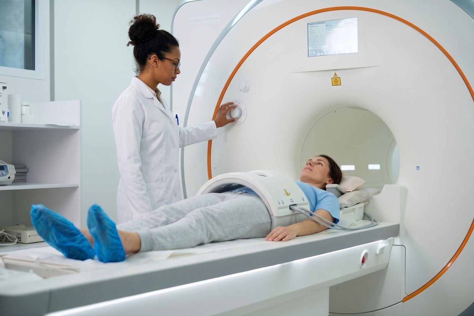
MRI scanners are noisy, so you may be given ear plugs or music. York explains that it is important to lie very still to stop the images becoming blurred. Because MRI uses strong magnets, you will be asked about pacemakers and metal implants beforehand. If you are nervous about enclosed spaces, see our guide to MRI options for claustrophobia.
Crohn's disease MRI scan vs normal: what the scan shows
People often search for a comparison of a Crohn's MRI and a normal scan. In simple terms, a radiologist looks at the bowel and the tissues around it and compares them with how they would normally appear. Findings the report may describe include:
- inflammation in the small bowel
- thickening of the bowel wall
- narrowed areas or blockages
- signs of bleeding
- fistulas, which are abnormal channels between the bowel and another area
The report also describes where any changes are and how much of the bowel seems to be involved. A report that does not show active inflammation is useful for your team, but it does not by itself mean Crohn's has gone away or that your symptoms should be ignored. Scans are read alongside your symptoms, blood and poo tests and camera tests. Royal Berkshire explains that the report is sent to the doctor who referred you. Our guide to how long scan results take has more on what happens next.
Pelvic MRI for perianal fistula
Crohn's can also affect the area around the bottom. The NHS lists Crohn's disease as one of the less common causes of an anal fistula, an abnormal channel near the anus. Symptoms can include skin irritation, a constant throbbing pain that may be worse when sitting, a smelly discharge, and passing pus or blood when you poo.
Tests can include scans such as ultrasound, CT or MRI. A pelvis MRI scan is commonly used. York and Scarborough say MRI is particularly effective in detecting fistulas, and a Health Research Authority research summary describes pelvic MRI as having an established role in assessing perianal fistula, helping doctors make clinical decisions and assess changes over time.
A fistula MRI is a different scan from MR enterography. It focuses on the pelvis and the area around the back passage, so your team will tell you how to prepare for it.
Where a private scan fits
MR enterography and fistula MRI are usually arranged by your IBD team as part of your ongoing care. Some people choose to have a scan privately, for example when their specialist has requested one. Not every clinic offers MR enterography, because it needs the drink, injections and a specific way of scanning.
If you are considering it, ask the clinic whether it performs MR enterography or fistula MRI, what referral it needs, and how the report and images will reach your IBD team so they can act on them. Our guide on getting your scan images and report may help.
Please speak to your GP or IBD team about any symptoms or results, and if symptoms are urgent, call NHS 111, or 999 in an emergency.
Finding a private scan
You can compare regulated clinics offering abdominal and pelvic MRI on FindScan, see each clinic's own listed price, and send an enquiry to the clinic you choose. The clinic will contact you to confirm whether it can do the scan you need and the final price. MRI does not use ionising radiation and some clinics accept self-referral, but for a specialist scan such as MR enterography it makes sense to have a request from your specialist, and the clinic will tell you what it needs.
Common questions
What is MR enterography?
MR enterography (MRE) is a special type of MRI scan that looks at the small bowel in detail. You drink a liquid beforehand to fill and expand the bowel, and contrast dye is usually injected during the scan. Crohn's and Colitis UK says it helps find inflammation, blockages and bleeding in the small bowel. It does not use X-rays.
How long does an MR enterography scan take?
The scan itself usually takes around 30 to 45 minutes, according to one NHS trust leaflet, but you should expect to be in the department for longer. You will need to arrive early to drink the bowel preparation, which can take about 45 minutes to fill the small bowel. In total you may be there for up to two hours.
Are there side effects from MR enterography?
The drink works as a mild laxative, so loose or watery bowel movements are common afterwards. If you have an injection of Buscopan to relax the bowel, it can cause a dry mouth and blurred vision for about 30 minutes, and you should not drive until your vision is normal. The team will explain the contrast dye and check it is suitable for you.
Is MRI or CT better for Crohn's disease?
Both have a role, and your IBD team will choose. MRI does not use X-rays, which is why one NHS trust says it is increasingly used for IBD, especially where repeated scans may be needed. CT scans involve a small dose of radiation. Ultrasound is another radiation-free option, and a study summarised by the NIHR found MRI slightly more accurate for the small bowel.
Can MRI show a Crohn's fistula?
Yes, MRI is often used for this. NHS guidance lists ultrasound, CT and MRI among the tests for an anal fistula, and one NHS trust says MRI is particularly effective in detecting fistulas. Pelvic MRI has an established role in assessing perianal fistula in Crohn's and in tracking changes over time. This is a different scan from MR enterography.
Can I eat before MR enterography?
Usually not for several hours. Instructions vary: one NHS trust asks people with morning appointments not to eat after midnight but to keep drinking water, while another asks for nothing to eat or drink for six hours. Always follow the instructions in your appointment letter, and tell the team about any regular medicines you take.
References
- Crohn's disease. NHS. Accessed 12 October 2026.
- Tests and investigations. Crohn's and Colitis UK. Accessed 12 October 2026.
- Having an MRI enterography (small bowel scan). Royal Berkshire NHS Foundation Trust. Accessed 12 October 2026.
- Small Bowel Magnetic Resonance Imaging (MRI) Enterography. Liverpool University Hospitals NHS Foundation Trust. Accessed 12 October 2026.
- Investigations and tests (Inflammatory bowel disease). York and Scarborough Teaching Hospitals NHS Foundation Trust. Accessed 12 October 2026.
- MRI and ultrasound scans are both helpful in assessing Crohn's disease; MRI is slightly more accurate. NIHR Evidence. Accessed 12 October 2026.
- Anal fistula. NHS. Accessed 12 October 2026.
- A novel MRI scoring system for Crohn's perianal fistula. Health Research Authority (NHS). Accessed 12 October 2026.
This article is general information, not medical advice, and it does not replace an assessment by a clinician. If you have symptoms that worry you, speak to your GP or call 111. In an emergency, call 999.