X-ray vs CT vs MRI: what is the difference?
X-ray, CT and MRI all take pictures of the inside of your body, but they work in different ways and suit different questions. This guide explains how each one works, what it is best at, how long it takes and how the radiation compares.
Written by the FindScan Editorial Team and reviewed by our Medical Advisor, Dr Mohamed Najjar
Reviewed
The quick comparison
X-ray, CT and MRI are the three imaging tests people ask about most. Here is how they differ at a glance:
- X-ray: uses a small amount of radiation to take a picture. It is mainly used for bones, teeth and lungs, and the X-ray itself takes only a few minutes.
- CT: uses X-rays and a computer to build up detailed cross-sectional pictures, or slices. It usually takes 10 to 20 minutes and gives a higher radiation dose than a plain X-ray.
- MRI: uses a magnetic field and radio waves, not X-rays. It shows soft tissues in detail and usually takes 15 minutes to 1 hour.
If you have been referred for a private X-ray, or are trying to understand why your clinician has suggested one test rather than another, the sections below explain each one in more detail.
How an X-ray works
The NHS describes an X-ray as a test that takes pictures of the inside of your body using a small amount of radiation. A standard X-ray is used mainly to check bones, teeth and lungs.
The picture works because different tissues let different amounts of X-ray through. Dense tissue such as bone allows few of the X-rays to pass through and appears white on the X-ray, while soft tissue lets more through and looks darker. This is why X-rays are so good at showing broken bones and changes in the joints, and why they are often the first test for these problems.
X-rays are quick. The NHS says having the X-ray itself should only take a few minutes. Common examples include a chest X-ray and X-rays of joints such as the knee or hip. Our guide to what a chest X-ray can show gives one example in more detail.
How a CT scan works
A CT scanner is a large ring that moves around part of your body as you lie on a flat bed. One NHS trust explains that it is a special X-ray machine which produces an image of a cross-section, or slice, of the body. A narrow beam of X-rays rotates around you, and a computer turns the information into detailed pictures of the inside of the body.
The NHS says a CT scan usually takes between 10 and 20 minutes. It lists uses such as checking bones or organs after an injury, finding the cause of symptoms, checking how well treatment is working and guiding procedures such as biopsies. Sometimes a dye called contrast medium is used to show certain areas more clearly. Our guide to CT scans with contrast explains what that involves, and our private CT scan page lists the types of CT scan offered.
How an MRI scan works
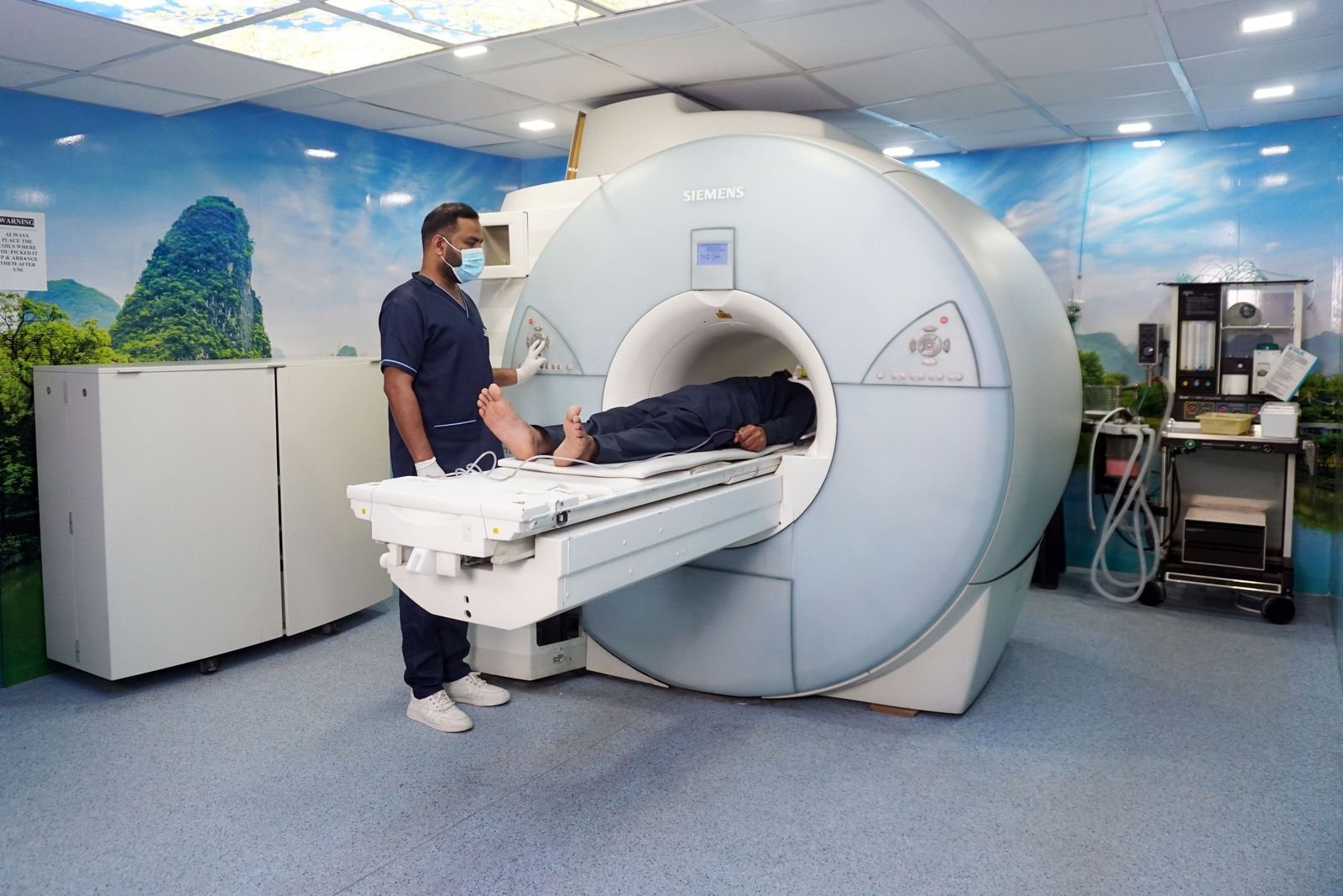
MRI stands for magnetic resonance imaging. One NHS leaflet explains that it works by using a magnetic field and radio waves, together with an advanced computer system, and that it does not involve the use of X-rays.
MRI is especially useful for soft tissues. For example, Versus Arthritis explains that an MRI of the knee will show the soft tissues, such as cartilage, tendons and muscles, and changes in the bone that cannot be seen on a standard X-ray.
The NHS says an MRI scan usually takes between 15 minutes and 1 hour, and the scanner makes loud bangs and clicks while it takes pictures. Because MRI uses a strong magnet, you will be asked about any metal in your body, such as surgical clips, a pacemaker or dental work, before the scan. See our guides to private MRI scans and MRI safety and metal implants.
CT vs X-ray: the key difference
CT and X-ray both use X-rays, which is why people often confuse them. The difference is in how the pictures are made and how much detail they show.
- Pictures: a standard X-ray takes a picture through the part of the body being examined. A CT scanner rotates around you and builds up many cross-sectional slices, which a radiologist can look through one by one.
- Detail: because CT produces slices, it can show organs and structures in more detail than a plain X-ray.
- Radiation: the trust leaflet above says that with CT the amount of radiation is more than is used for an ordinary X-ray of the chest or body.
- Time: an X-ray takes a few minutes, while a CT scan usually takes 10 to 20 minutes.
A plain X-ray is often the first step, for example for a suspected fracture. CT may be used when more detail is needed, depending on the question your clinician is trying to answer.
X-ray vs MRI
X-ray and MRI are very different tests:
- X-ray uses a small amount of radiation, while MRI uses a magnetic field and radio waves.
- X-ray is best for bone, while MRI shows soft tissues such as cartilage, tendons and muscles.
- X-ray takes a few minutes, while MRI usually takes 15 minutes to 1 hour.
A good example is knee pain. Versus Arthritis says X-rays are the most useful tests to confirm a diagnosis of osteoarthritis, although they won't often be needed. MRI may be suggested to look at soft tissues, but this is less common. Neither test is better in every situation. They answer different questions.
Radiation: how the doses compare
Radiation dose is measured in millisieverts (mSv). The UK Health Security Agency (UKHSA) publishes typical doses for common X-ray and CT examinations, alongside the equivalent amount of natural background radiation that we all receive from the environment:
- Chest X-ray: 0.02 mSv, about 3 days of natural background radiation
- X-ray of limbs and joints (except the hip): less than 0.01 mSv, less than 1.5 days
- CT head: 2 mSv, about 1 year
- CT chest: 8 mSv, about 3.6 years
- CT abdomen and pelvis: 10 mSv, about 4.5 years
UKHSA puts the UK average background dose at 2.1 mSv a year. MRI does not use X-rays, so it does not involve this kind of radiation dose.
These figures can look large, but they need context. The NHS says that if you have lots of CT scans, there's a very small chance the radiation from the X-rays can cause cancer, and that doctors weigh up the risks before giving you a scan. This is why every X-ray and CT scan has to be justified before it goes ahead, as explained below. Our guide to X-ray radiation safety explains more.
Why the referrer chooses the test
You might wonder whether you can simply pick the scan you prefer. In practice, the right test depends on your symptoms and the question that needs answering, and that is a clinical decision.
For X-ray and CT, the law also has a say. Under the Ionising Radiation (Medical Exposure) Regulations 2017 (IR(ME)R), a person must not carry out an exposure unless it has been justified by the practitioner as showing a sufficient net benefit. In practice, every X-ray and CT scan needs a referral from a referrer, such as a GP or consultant, who must give the imaging service enough medical information to judge whether the scan is worthwhile. The practitioner at the imaging service then decides whether the expected benefit is enough to justify the exposure.
MRI does not use ionising radiation, so IR(ME)R does not apply in the same way. Even so, a clinician still needs to decide that MRI is the right test, and the clinic will check that it is safe for you. If you are weighing up MRI against CT in particular, our guide to MRI vs CT scans goes into more detail.
This article is general information, not medical advice. Please speak to your GP or the clinician who referred you about your symptoms or results, and if your symptoms are urgent, call NHS 111 or 999.
Finding a private scan
You can compare private X-ray, CT and MRI clinics on FindScan, see each clinic's own listed price and send an enquiry to the clinic you choose. The clinic will contact you to confirm the details and the final price. X-ray and CT scans always need a referral under IR(ME)R, and many private clinics can arrange this through their own doctor. Some clinics accept self-referral for MRI, depending on the clinic.
Common questions
What is the difference between a CT scan and an X-ray?
Both use X-rays. A standard X-ray takes a picture through the part of the body being examined and takes a few minutes. A CT scanner rotates around you and uses a computer to build up detailed cross-sectional slices. CT usually takes 10 to 20 minutes, shows more detail and gives a higher radiation dose than a plain X-ray.
Is MRI better than CT or X-ray?
No single scan is best for everything. X-ray is quick and good for bones and lungs. CT gives detailed slices of the body and is quick. MRI shows soft tissues such as cartilage, tendons and muscles in detail and does not use X-rays, but takes longer. The right test depends on the question your clinician needs to answer.
Which scan gives the most radiation?
Of the three, CT usually gives the highest dose. UKHSA figures put a CT of the abdomen and pelvis at about 10 mSv, compared with 0.02 mSv for a chest X-ray. MRI does not use X-rays. Doctors weigh up the risks and benefits before any X-ray or CT scan, as the law requires.
Which scan is quickest?
A plain X-ray is quickest. The NHS says the X-ray itself only takes a few minutes. A CT scan usually takes 10 to 20 minutes, and an MRI scan usually takes between 15 minutes and 1 hour. You should also allow time for checking in, safety questions and changing, which adds to the total appointment time.
Can I choose which scan I have?
The choice of test is a clinical decision based on your symptoms. For X-ray and CT, the Ionising Radiation (Medical Exposure) Regulations 2017 require a referrer, and the exposure must be justified as having a sufficient net benefit. You can discuss the options with your clinician, who can explain why one test suits your situation better than another.
References
- X-ray. NHS (nhs.uk). Accessed 12 October 2026.
- CT scan. NHS (nhs.uk). Accessed 12 October 2026.
- MRI scan. NHS (nhs.uk). Accessed 12 October 2026.
- X-Rays. Blackpool Teaching Hospitals NHS Foundation Trust. Accessed 12 October 2026.
- Your Computed Tomography (CT) Examination. Doncaster and Bassetlaw Teaching Hospitals NHS Foundation Trust. Accessed 12 October 2026.
- Adults having an MRI scan. Gloucestershire Hospitals NHS Foundation Trust. Accessed 12 October 2026.
- Patient dose information: guidance. GOV.UK (UK Health Security Agency). Accessed 12 October 2026.
- Osteoarthritis of the knee. Versus Arthritis. Accessed 12 October 2026.
- The Ionising Radiation (Medical Exposure) Regulations 2017, regulation 11. legislation.gov.uk. Accessed 12 October 2026.
- The Ionising Radiation (Medical Exposure) Regulations 2017, regulation 10. legislation.gov.uk. Accessed 12 October 2026.
This article is general information, not medical advice, and it does not replace an assessment by a clinician. If you have symptoms that worry you, speak to your GP or call 111. In an emergency, call 999.