Ultrasound vs MRI: When Ultrasound Is Enough
Ultrasound is quicker, cheaper, radiation-free and sometimes the better test. MRI sees deeper and in more detail. Here is where the line falls, body part by body part.
Written by the FindScan Editorial Team and reviewed by our Medical Advisor, Dr Mohamed Najjar
Reviewed
Ultrasound and MRI overlap more than people expect. Both use no ionising radiation, both are good at soft tissue, and for a surprising number of questions, a shoulder tendon, a lump under the skin, the gallbladder, the thyroid, ultrasound is the first-line test and often the only one needed. MRI earns its longer appointment and higher cost when the problem is deep, complex, inside a joint or inside the skull or spine, or when ultrasound has already been done and left a question open. This guide sets out what each does well, where each falls short, and how the decision is usually made in UK practice.
This page is general information, not medical advice. If you have symptoms that worry you, speak to your GP.
How each scan works, briefly
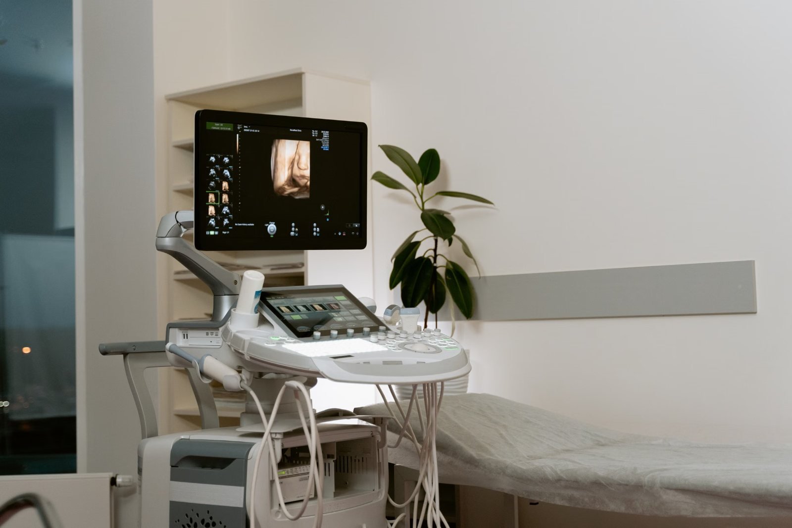
Ultrasound sends high-frequency sound into the body through a hand-held probe and builds an image from the echoes. It is live, so the operator can watch a tendon move or a joint flex, and it can show blood flow with Doppler. Its limits are physical: sound does not pass through bone or gas, so it cannot see inside the skull, behind a rib, or through a gassy bowel, and detail falls off with depth, which matters for larger patients and deep structures.
MRI uses a strong magnet and radio waves to map the hydrogen in tissue. It sees through bone and gas, images a whole region in one sitting rather than wherever the probe is pointed, and gives a permanent, reviewable set of slices rather than a snapshot the operator chose. It is also slower, noisier, more enclosed and more expensive, and it has its own safety rules around implants. The NHS says an ultrasound typically takes around 10 to 45 minutes and an MRI between 15 minutes and an hour, though both can take longer.
Joints, tendons and muscles
This is the most common place the two compete. Ultrasound is excellent for structures near the surface: the rotator cuff tendons of the shoulder, the Achilles tendon, tennis elbow, plantar fascia, a suspected muscle tear, and fluid in or around a joint. It can be done quickly, compared with the other side, and used to guide an injection in the same visit.
MRI takes over for anything inside the joint. Cartilage, the menisci and cruciate ligaments of the knee, the labrum of the hip or shoulder, bone bruising, stress fractures and early arthritis changes are MRI territory. A good rule of thumb: if the structure sits on the outside of a joint and can be reached by a probe, ask whether ultrasound would do; if the problem is within the joint, expect MRI. Our guides to shoulder MRI and knee MRI go into what each shows.
Abdomen and pelvis
Ultrasound is the standard first test for the gallbladder, liver, kidneys, the thyroid, the testes, and the female pelvis, where transvaginal ultrasound often gives better pictures than anything else. It is also the NHS screening tool for abdominal aortic aneurysm. The NHS lists investigating pain or a lump and monitoring a condition such as a thyroid problem among the main reasons for an ultrasound.
MRI is used when ultrasound cannot see what is needed, for example to characterise a liver lesion, to assess the bile ducts or pancreas, for endometriosis mapping, for fibroids before treatment, and for the prostate, where multiparametric MRI has replaced ultrasound-guided biopsy as the first step in the UK pathway. Our article on what an abdominal ultrasound shows covers the ultrasound side in detail.
Lumps, bumps and soft tissue
A lump you can feel is almost always an ultrasound question first. Ultrasound will tell the difference between a cyst and a solid lump, show whether a lymph node looks reactive or worrying, and guide a needle if a sample is needed. MRI is reserved for lumps that are deep, large, fixed, or that ultrasound has flagged as needing more detail before surgery. For breast symptoms the pathway is different again and is led by the NHS breast clinic, where ultrasound and mammography sit together.
Where ultrasound cannot help
Some questions are simply not ultrasound questions. The brain and spinal cord are behind bone; back pain and sciatica need MRI if imaging is indicated at all; the lungs are full of air; and most of the bowel is obscured by gas. If a clinic offers ultrasound for a headache, persistent back pain or a suspected disc problem, that is a reason to be cautious. Our guide to which scan for back pain and sciatica explains why, and the NHS pages on MRI and ultrasound set out what each is used for.
Operator skill, and why it matters more for ultrasound
An MRI produces the same set of images whoever presses the button, within reason. Ultrasound does not: the quality of the study depends on the person holding the probe and on what they chose to look at. That is why ultrasound reports often describe what was examined as well as what was found, and why a negative ultrasound of a joint from an experienced musculoskeletal sonographer or radiologist carries more weight than one from a general list. When you enquire with a clinic, it is reasonable to ask who performs and who reports the ultrasound, and whether they specialise in that area of the body. The report should be written by an appropriately qualified clinician for that type of imaging.
Cost, access and referral
Ultrasound is usually the cheaper of the two, the quicker to arrange, and the scan more likely to be reported on the day. Neither ultrasound nor MRI uses ionising radiation, so neither carries the legal referral requirement that applies to X-ray and CT, and many private clinics accept self-referral for both. The practical sequence for a lot of problems is ultrasound first, MRI only if it is still needed, which is cheaper and faster than going straight to MRI and often answers the question outright. If a clinic suggests the opposite, ask them to explain why, and expect a reason tied to your symptoms. Our comparison of MRI and CT covers the other common fork in the road.
Common questions
Is MRI better than ultrasound?
Not in general. Ultrasound is better for surface tendons, lumps, the gallbladder, thyroid and much of the pelvis, and it is live. MRI is better for structures inside joints, the brain and spine, and deep organs. The right test depends on the question.
Can ultrasound see a torn ligament?
Some, such as the ankle ligaments and the collateral ligaments of the knee, can be assessed with ultrasound. Ligaments inside a joint, such as the cruciate ligaments, need MRI.
Do I need a referral for an ultrasound or an MRI?
Neither uses ionising radiation, so there is no legal referral requirement as there is for X-ray and CT. Many private clinics accept self-referral for both, though some ask for a clinician's request for certain studies.
Should I have an ultrasound before an MRI?
For many surface and abdominal problems, yes: ultrasound is quicker and cheaper and often settles the question. For joint interiors, the spine and the brain, going straight to MRI is usual.
References
- Ultrasound scan: why it is done, what happens and getting your results. NHS. Accessed 5 October 2026.
- MRI scan: why it is done and what happens. NHS. Accessed 5 October 2026.
- Abdominal aortic aneurysm screening. NHS. Accessed 5 October 2026.
- The Ionising Radiation (Medical Exposure) Regulations 2017, SI 2017/1322 (legislation.gov.uk). legislation.gov.uk. Accessed 5 October 2026.
- iRefer: making the best use of clinical radiology. Royal College of Radiologists. Accessed 5 October 2026.
This article is general information, not medical advice, and it does not replace an assessment by a clinician. If you have symptoms that worry you, speak to your GP or call 111. In an emergency, call 999.