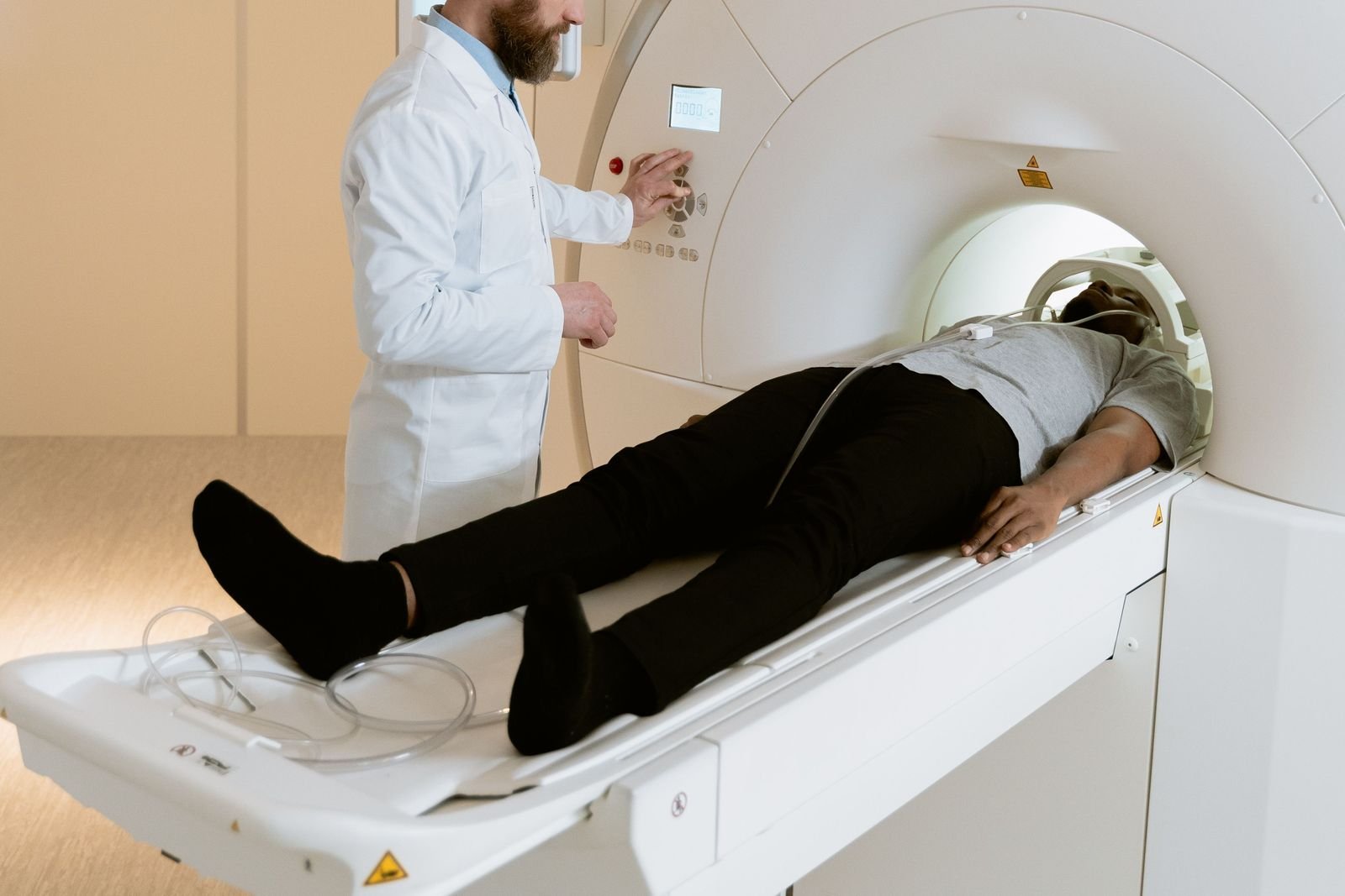
MRI scan of the leg and thigh: what it shows and when it helps
An MRI scan of the leg can give a detailed look at muscles, tendons and bone when a thigh or calf problem is not settling. This guide explains what leg MRI shows, when ultrasound is used first, and which symptoms need urgent NHS care instead.
Written by the FindScan Editorial Team and reviewed by our Medical Advisor, Dr Mohamed Najjar
Reviewed
What a leg or thigh MRI can show
An MRI scan of the leg uses a strong magnet and radio waves to make detailed pictures of the muscles, tendons, other soft tissues and bones. You can compare clinics offering a private MRI scan of the thigh, calf or other areas on FindScan.
Common reasons for a leg MRI include:
- muscle and tendon injuries, such as hamstring and calf tears
- suspected stress fractures that do not show on an X-ray
- soft tissue lumps that need a closer look after an initial assessment
If your pain is centred on a joint, a scan of that joint is usually more useful than a general leg scan. See our pages on knee MRI, hip MRI and foot and ankle MRI.
Hamstring injuries
The hamstrings are the group of muscles and tendons at the back of the thigh. The NHS describes hamstring injury symptoms as a sharp pain at the back of the leg, difficulty standing or walking, and tenderness, swelling or bruising after a few hours. Mild injuries usually get better in a few days, but severe injuries can take weeks or months to heal and may need specialist treatment.
Many hamstring injuries do not need a scan. An NHS physiotherapy leaflet on hamstring strains from Essex Partnership University NHS Foundation Trust says diagnosis is usually made from your history and an examination. An ultrasound or MRI may be needed if a more severe injury is suspected, or if recovery is not going as expected.
Hamstring injuries are often graded from 1, a mild strain, to 3, a complete tear. If the hamstring tendons have pulled away from the bone near the buttock, the leaflet says you are likely to be referred to an orthopaedic surgeon to discuss repair. In these situations, a hamstring MRI can help show where the injury is and how extensive it is.
Calf injuries
Calf strains are managed in a similar way. The same trust's calf strain leaflet says diagnosis is generally made from your history and examination, with an ultrasound or MRI if a more severe injury is suspected or symptoms are not improving as expected.
Pain and swelling in the calf can also be caused by a blood clot, which needs a different and urgent route. This is covered in the red flags section below.
Stress fractures
A stress fracture is a small crack in a bone that builds up from repeated strain rather than one single injury. These can be hard to see at first. RCEMLearning, from the Royal College of Emergency Medicine, notes that the first X-ray is frequently normal. It lists MRI, a bone scan, or a repeat X-ray after 10 days as ways to confirm the diagnosis.
Your clinician will decide which test makes sense for you. X-rays and bone scans use ionising radiation, so they always need a referral from a clinician. MRI does not use ionising radiation.
Soft tissue lumps
Lumps in the leg or thigh can have many causes, but some need to be checked promptly. Sarcoma UK lists warning signs that should be seen by a GP:
- a lump or swelling of any size that is getting bigger or changing
- a lump larger than 5 cm, about the size of a golf ball
- a lump that feels deep under the skin
- pain from a lump or swelling
Sarcoma UK adds that these symptoms can have many other causes and do not mean you have sarcoma.
The first test is often not MRI. Sarcoma UK says your GP may arrange an ultrasound or X-ray first. NICE guidance on suspected cancer (NG12), as summarised by Macmillan Cancer Support, says GPs should consider an urgent ultrasound, within two weeks, for adults with an unexplained lump that is increasing in size. It also advises an urgent referral if the ultrasound suggests a sarcoma, or if the findings are uncertain and concern remains. After referral, the NHS explains that a specialist may arrange further tests, including ultrasound, CT and MRI scans and a biopsy.
This is why a growing lump should go through your GP rather than straight to a private scan. The NHS pathway is set up to get people with worrying lumps seen quickly by the right specialists.
When ultrasound is used first
Ultrasound is often the first scan for leg problems. It does not use radiation, it is the scan the NHS uses to check for a blood clot in the leg, and it is commonly used first for lumps and for some muscle injuries. You can read about the differences in our guide to ultrasound vs MRI, or compare clinics offering a private ultrasound scan.
MRI is more likely to be suggested when the picture is still unclear, the injury seems severe, a stress fracture is suspected, or a specialist needs more detail to plan treatment.
Red flags: when you need urgent NHS care
Do not wait for a private scan if you have any of the following:
- A possible DVT (blood clot). The NHS lists DVT symptoms as throbbing pain in one leg, usually in the calf or thigh, swelling, red, blue or darkened skin around the painful area, and swollen veins. Ask for an urgent GP appointment or contact NHS 111. The NHS says you should be referred to hospital within 24 hours for an ultrasound scan.
- DVT symptoms with breathlessness or chest pain. Call 999 or go to A&E. The NHS warns that a pulmonary embolism, a clot in the lungs, can be life-threatening and needs treatment straight away.
- Tingling, loss of feeling or coldness in the lower leg or foot after an injury. The EPUT calf strain leaflet says this needs assessment in the Emergency Department.
- A lump that is growing quickly or changing. See your GP promptly so they can arrange the right tests, which may include an urgent referral.
Why a clinician's referral letter helps
The leg is long, and an MRI scan is planned around a specific area. The radiographer needs to know where to centre the scan and what question it needs to answer. A short letter from your GP, physiotherapist or sports medicine doctor can make the scan much more useful. Helpful details include:
- exactly where the pain or lump is, for example the back of the upper thigh
- how and when the injury happened
- what the clinician found on examination and what they suspect
- any previous X-rays, ultrasound scans or operations
Some clinics accept self-referral for MRI, but even then, bringing this information helps target the scan. It also means there is a clinician ready to act on the report. See our guide to private MRI without a GP referral for more.
What to expect on the day
The NHS says an MRI scan usually takes between 15 minutes and 1 hour, and can take longer. The scanner makes loud bangs and clicks, so you will be offered ear plugs or headphones, and you will need to lie still. You will be asked about any metal in your body beforehand, and you may sometimes have an injection of contrast dye to show more detail. You can eat and drink as normal afterwards.
For more detail, read what to expect at an MRI scan and our guide to MRI safety and metal implants.
Please speak to your GP, or the clinician who referred you, about any symptoms or results, and if symptoms are urgent, call NHS 111, or 999 in an emergency.
Finding a private scan
You can compare regulated clinics offering leg and thigh MRI on FindScan, see each clinic's own listed price, and send an enquiry to the clinic you choose. The clinic will contact you to confirm the details and the final price. MRI does not use ionising radiation and some clinics accept self-referral while others ask for a referral, so check with the clinic. If you also need an X-ray, that always needs a referral from a clinician, which many private clinics can arrange through their own doctor.
Common questions
What does an MRI scan of the leg show?
A leg or thigh MRI shows muscles, tendons, other soft tissues and bone in detail. It is often used to look at more severe muscle and tendon injuries, such as hamstring or calf tears, suspected stress fractures that are not visible on X-ray, and soft tissue lumps after an initial assessment. If the problem is in a joint, a joint scan is usually better.
Do I need an MRI for a hamstring injury?
Often not. NHS physiotherapy guidance says hamstring injuries are usually diagnosed from your history and an examination. An ultrasound or MRI may be needed if a more severe injury is suspected, if recovery is slower than expected, or if the tendon may have pulled away from the bone, which can lead to a referral to an orthopaedic surgeon.
Is ultrasound or MRI better for a thigh or calf injury?
It depends on the question. Ultrasound is quick, does not use radiation and is often used first. MRI gives a broader, detailed view and is more likely to be suggested when an injury seems severe, is not improving, or a stress fracture is suspected. NHS leaflets for hamstring and calf strains mention both as options when further investigation is needed.
Can a private MRI check for a blood clot in my leg?
If you think you might have a DVT, you need urgent NHS care, not a private MRI. The NHS advises an urgent GP appointment or NHS 111, and says you should be referred to hospital within 24 hours for an ultrasound scan. If you also feel short of breath or have chest pain, call 999 or go to A&E.
Should I get a private MRI for a lump in my thigh?
See your GP first. Sarcoma UK says a lump that is getting bigger, larger than about 5 cm, deep under the skin or painful should be checked, and your GP may arrange an ultrasound or X-ray first. NICE guidance supports an urgent ultrasound for an unexplained lump that is increasing in size, with urgent specialist referral if needed.
How long does a leg MRI scan take?
The NHS says MRI scans usually take between 15 minutes and 1 hour, and can take longer. The exact time depends on the area being scanned and whether contrast dye is needed. You will need to lie still, the scanner is noisy so you will be offered ear protection, and you can eat and drink as normal afterwards.
References
- Hamstring injury. NHS. Accessed 12 October 2026.
- Hamstring Strains (patient information leaflet). Essex Partnership University NHS Foundation Trust. Accessed 12 October 2026.
- Calf strains (patient information leaflet). Essex Partnership University NHS Foundation Trust. Accessed 12 October 2026.
- Confirming a Stress Fracture. RCEMLearning (Royal College of Emergency Medicine). Accessed 12 October 2026.
- Signs and symptoms of sarcoma (Do I have sarcoma?). Sarcoma UK. Accessed 12 October 2026.
- Rapid Referral Guidelines for Bone Cancer and Sarcoma. Macmillan Cancer Support (summary of NICE NG12). Accessed 12 October 2026.
- Tests and next steps for soft tissue sarcoma. NHS. Accessed 12 October 2026.
- DVT (deep vein thrombosis). NHS. Accessed 12 October 2026.
- MRI scan. NHS. Accessed 12 October 2026.
This article is general information, not medical advice, and it does not replace an assessment by a clinician. If you have symptoms that worry you, speak to your GP or call 111. In an emergency, call 999.