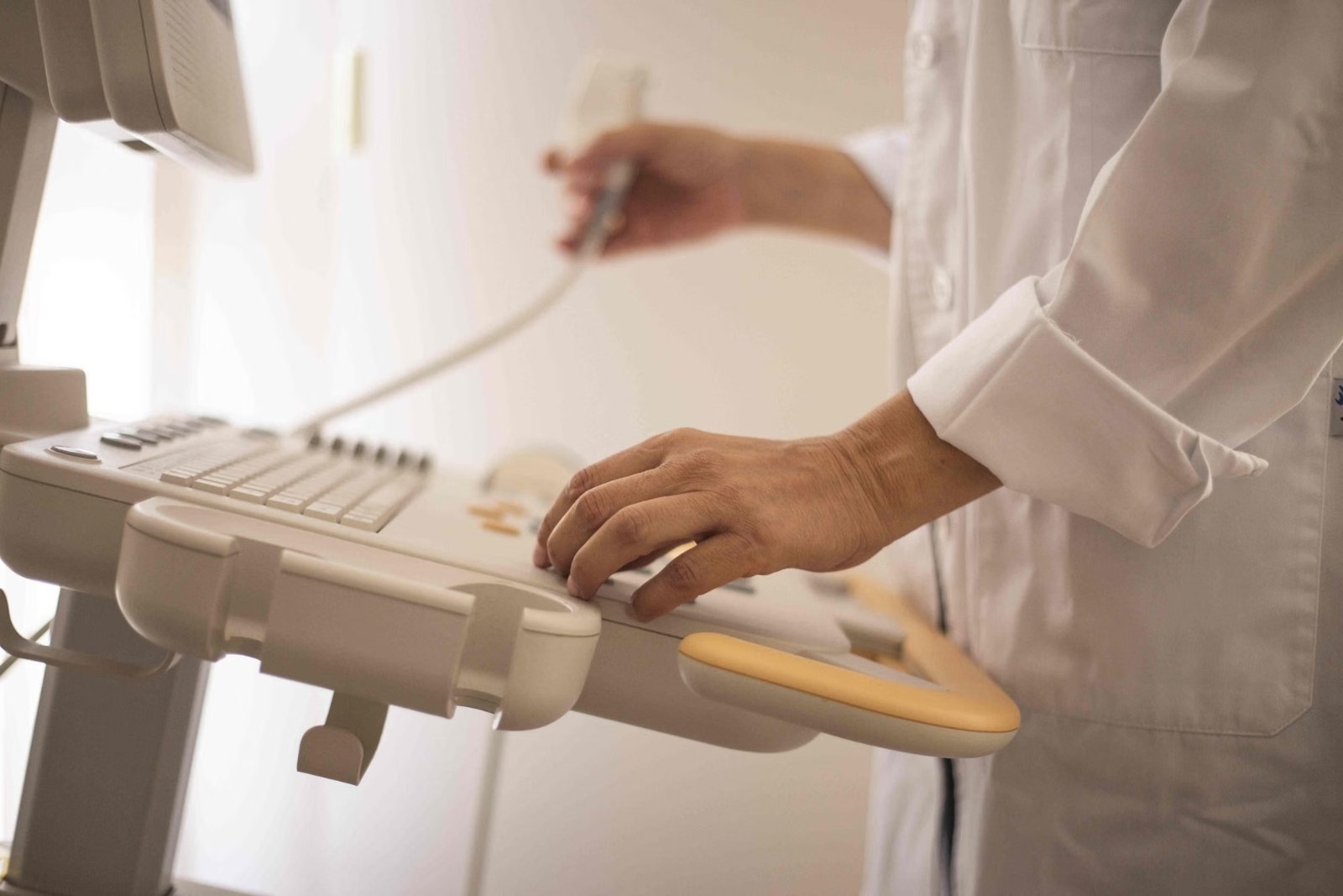
Types of echocardiogram: transthoracic, TOE, bubble and stress echo
Most echocardiograms are transthoracic, with a probe moved over the chest, but there are other types for different situations. Here is what each type involves, why it is used and how to prepare.
Written by the FindScan Editorial Team and reviewed by our Medical Advisor, Dr Mohamed Najjar
Reviewed
An echocardiogram, or echo, is an ultrasound scan of the heart. Most people who have one will have a transthoracic echocardiogram, where a probe is moved over the chest. But there are other types too, including the transoesophageal echo, the bubble echo and the stress echo. Each is used for a different reason and feels quite different for the patient. This guide explains what each involves, why it is used and how to prepare. If you are looking into a private echo, you can compare private echocardiogram clinics on FindScan.
A quick overview
The NHS guide to echocardiograms describes the main types:
- Transthoracic echocardiogram (TTE): the most common type, with the probe moved over the chest.
- Stress echo: similar to a TTE, but with scans taken before and after your heart is made to work harder.
- Transoesophageal echocardiogram (TOE): a thin tube with a small ultrasound probe is passed down the throat into the food pipe for more detailed pictures.
The British Heart Foundation also describes contrast echo, bubble echo and foetal echo, which is used to look for heart problems in a baby before birth.
Transthoracic echocardiogram (TTE)
What it is and why it is used
A transthoracic echocardiogram is the standard echo. "Transthoracic" simply means "through the chest". The NHS says an echo can be used to check whether symptoms may be caused by a heart condition, to monitor people with a known heart problem, and to help diagnose conditions such as heart failure, heart valve disease and congenital heart disease. It looks at the structure of the heart, including the valves, and how well it pumps.
What happens
You undress from the waist up and wear a gown, then lie on a couch. In its patient leaflet on echocardiograms, University Hospital Southampton explains that you usually lie on your left side, the lights are dimmed so the sonographer can see the screen, and a probe covered in gel is placed on your chest. Small sticky patches are usually attached to monitor your heart rate. The machine may make a loud swishing sound, which is normal.
How long it takes and how it feels
The NHS says a TTE takes about 30 to 40 minutes. The Southampton leaflet describes it as a completely painless test with no side effects that does not use radioactivity. The gel feels cold, and you may feel some pressure from the probe. For more on timings, see our guide on how long an echocardiogram takes.
Preparation
You can usually eat, drink and take your medicines as normal. Wear clothing that is easy to remove from the top half. You can ask for a chaperone. A TTE is widely available, both in hospitals and in many community and private clinics.
Contrast echo
Sometimes the pictures from a standard echo are not clear enough. The NHS explains that a contrast agent may be injected into a vein in your arm during a TTE to make the heart clearer. If you have contrast, you may be asked to wait up to 30 minutes afterwards so staff can check for any reaction.
Bubble echocardiogram
What it is and why it is used
A bubble echocardiogram is a TTE with an injection of tiny bubbles made from salt water. The bubbles show up clearly on the scan, which helps the sonographer look for a small hole in the heart. The BHF says it can help identify a hole in the heart and may be done after a stroke, a mini stroke (TIA) or complex heart surgery.
The Gloucestershire Hospitals bubble echo leaflet explains that a common reason is a suspected patent foramen ovale (PFO), a small flap-like channel between the upper chambers of the heart.
What happens
A small plastic tube called a cannula is placed in a vein in your arm or hand. The bubbles are made by quickly mixing sterile salt water with a little air and some of your blood, then injected through the cannula while the heart is scanned. You may be asked to sniff or to do a special breathing technique called the Valsalva manoeuvre, a special breathing and blowing technique. Staff will explain it and let you practise first.
How it feels, timing and preparation
The Gloucestershire leaflet says the test takes about 30 to 45 minutes and that complications are rare. The breathing technique may make your ears pop or cause a slight headache. You can usually eat, drink and take your medicines as normal, and staying well hydrated with warm hands can make the cannula easier to place.
Stress echocardiogram
What it is and why it is used
A stress echocardiogram looks at how your heart behaves when it is working harder. Some heart problems only show up during exertion. The Royal Free London stress echo leaflet says it can help find out whether symptoms such as angina or breathlessness are due to underlying heart disease. The BHF adds that it can help diagnose coronary heart disease, which is caused by narrowing of the arteries that supply the heart muscle.
What happens
Pictures are taken at rest and then again when your heart rate is raised. This is done in one of two ways:
- Exercise: on a treadmill or exercise bike.
- Medicine: usually a drug called dobutamine, given as an injection into a vein, which makes the heart beat faster and harder, similar to exercise.
Sticky electrodes on your chest monitor your heart throughout the test.
How long it takes and risks
The Royal Free leaflet says a stress echo takes about 45 to 60 minutes. It describes the test as very safe, but with some small risks, including an abnormal heart rhythm in about 1 in 1,000 patients, which usually settles without treatment. It advises not driving after a dobutamine test for the rest of the day.
Preparation
Preparation is more involved than for a standard echo. You may be asked to stop rate controlling medicines, such as beta blockers, for up to 48 hours beforehand, but only if your doctor tells you to. The Royal Free asks patients to avoid caffeine for 24 hours, and the Gloucestershire stress echo leaflet asks patients not to eat for 2 hours before. Instructions vary between hospitals, so always follow the ones you are given. Wear comfortable clothes and shoes if you might be exercising.
Transoesophageal echocardiogram (TOE)
What it is and why it is used
A transoesophageal echocardiogram, sometimes spelt transesophageal and known in the US as a TEE, places the ultrasound probe inside the food pipe (oesophagus), which sits just behind the heart. Because the probe is much closer, the University Hospital Southampton TOE leaflet says the pictures can be much more detailed than a standard echo. The Milton Keynes University Hospital TOE leaflet explains that a TOE is often used to look carefully at the valves and the structure of the heart, and to help the doctor decide on the best treatment.
What happens
A doctor sprays the back of your throat with local anaesthetic to numb it, and a mouth guard protects your teeth. A sedative is usually given through a cannula to help you relax. The NHS says you will not be asleep, but you will be very drowsy. The thin, flexible tube is then passed through your mouth into your food pipe.
How long it takes and how it feels
The Milton Keynes leaflet says the test itself takes about 20 minutes, with recovery usually taking 2 to 3 hours. It can feel unpleasant but should not be painful. A sore throat is common for a day or two afterwards. Serious complications are very rare: the Milton Keynes leaflet puts the risk of damaging the gullet at less than 1 in 10,000.
Preparation
You will usually be asked not to eat or drink for 6 hours before the test. If you take a blood thinner such as warfarin, you may need a blood test beforehand, so tell the team. Because of the sedation, you will need someone to take you home. The NHS advises not driving for 24 hours, and the Southampton leaflet asks that someone stays with you overnight.
Which types are hospital procedures?
A standard TTE, and often a bubble or contrast echo, can be done as an outpatient appointment. A TOE is typically a hospital procedure, carried out in a cardiology department, because it needs a doctor, sedation, a fasting period and a recovery area. A stress echo, especially one using dobutamine, is also typically done in a hospital cardiology department where staff can monitor you and respond to any problems. If a standard echo cannot give clear enough pictures, NICE guidance on heart failure notes that doctors may consider other imaging, such as a TOE or a cardiac MRI scan (NICE guideline NG106).
This article is general information, not medical advice. Please speak to your GP or the clinician who referred you about your symptoms or results, and if symptoms are urgent, call NHS 111, or 999 in an emergency.
Finding a private scan
You can compare private echocardiogram clinics on FindScan, where each clinic lists its own price and the types of echo it offers, and send an enquiry to the one you choose. The clinic will contact you to confirm the details. Because an echo uses ultrasound rather than radiation, some clinics accept self-referral for a standard transthoracic echo, while others ask for a referral from your GP or their own doctor; stress echo and TOE are usually arranged through a cardiologist.
Common questions
What is a transthoracic echocardiogram?
A transthoracic echocardiogram (TTE) is the standard, most common type of echo. A sonographer moves a probe with gel over your chest to create moving pictures of the heart's chambers and valves and to see how well it pumps. NHS information says it takes about 30 to 40 minutes, and it is painless and does not use radiation.
How is a transoesophageal echocardiogram different from a standard echo?
In a transoesophageal echo (TOE, or TEE in the US), the probe is passed down the throat into the food pipe, which sits just behind the heart. This gives more detailed pictures, especially of the valves. It involves a throat spray, usually sedation, fasting for about 6 hours beforehand and someone to take you home afterwards.
Why would I need a bubble echocardiogram?
A bubble echo is often used to check for a small hole between the upper chambers of the heart, such as a patent foramen ovale (PFO). The British Heart Foundation says it may be done after a stroke, a mini stroke (TIA) or complex heart surgery. Tiny saline bubbles are injected into an arm vein while the heart is scanned.
What should I avoid before a stress echocardiogram?
Instructions vary, so follow your hospital's advice. Some NHS leaflets ask patients to avoid caffeine for 24 hours and not to eat for a couple of hours beforehand. You may be asked to stop beta blockers or other rate controlling medicines for up to 48 hours, but only if your doctor tells you to. Do not stop any medicine without advice.
Can I drive after an echocardiogram?
After a standard transthoracic echo, you can usually carry on as normal. After a TOE with sedation, the NHS advises not driving for 24 hours, and you will need someone to take you home. After a dobutamine stress echo, the Royal Free advises not driving for the rest of the day. Always follow the advice you are given.
References
- Echocardiogram. NHS. Accessed 12 October 2026.
- Echocardiogram (echo). British Heart Foundation. Accessed 12 October 2026.
- Echocardiogram: patient information. University Hospital Southampton NHS Foundation Trust. Accessed 12 October 2026.
- Bubble contrast echocardiogram (echo). Gloucestershire Hospitals NHS Foundation Trust. Accessed 12 October 2026.
- Having a stress echocardiogram (echo). Royal Free London NHS Foundation Trust. Accessed 12 October 2026.
- Stress echocardiogram. Gloucestershire Hospitals NHS Foundation Trust. Accessed 12 October 2026.
- Transoesophageal echocardiogram (TOE): patient information. University Hospital Southampton NHS Foundation Trust. Accessed 12 October 2026.
- Transoesophageal echocardiogram (TOE): patient information leaflet. Milton Keynes University Hospital NHS Foundation Trust. Accessed 12 October 2026.
- Chronic heart failure in adults: diagnosis and management (NG106), Recommendations. NICE. Accessed 12 October 2026.
This article is general information, not medical advice, and it does not replace an assessment by a clinician. If you have symptoms that worry you, speak to your GP or call 111. In an emergency, call 999.