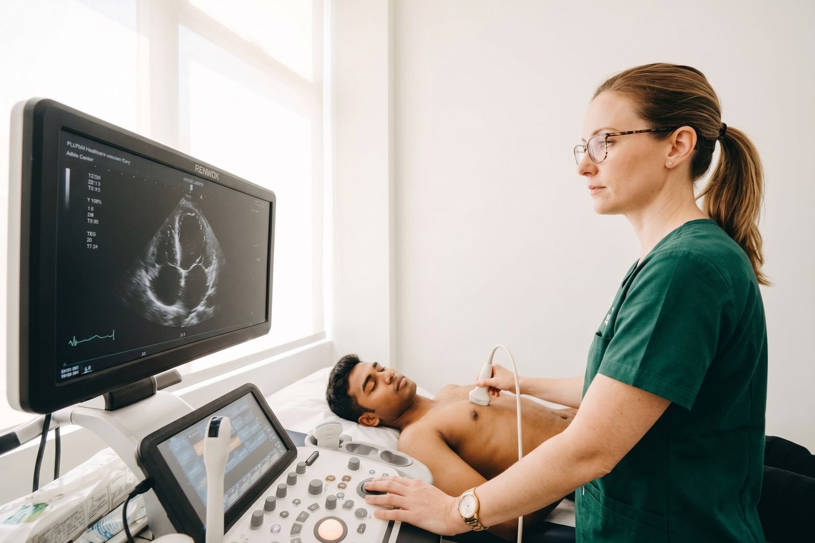
What will an echocardiogram show?
An echocardiogram shows your heart's chambers and valves and how well it pumps. Here is what it can show, what ejection fraction means, and what an echo cannot show well.
Written by the FindScan Editorial Team and reviewed by our Medical Advisor, Dr Mohamed Najjar
Reviewed
If you have been referred for a heart scan, it is natural to wonder what an echocardiogram will show. An echo is an ultrasound scan of the heart. It gives doctors moving pictures of the heart's chambers and valves and shows how well the heart is pumping. It is a very useful test, but it also has limits. This guide explains what it can and cannot show, in plain English. If you are considering a private scan, you can compare private echocardiogram clinics on FindScan.
How an echo works, in brief
A sonographer moves a small probe with gel over your chest. The probe sends out sound waves that bounce off the heart and are turned into live pictures on a screen. The British Heart Foundation explains that an echo looks at the structures of your heart, such as the heart valves, and gives information on how well your heart is pumping. It can also show blood flowing through the heart's chambers. For timings, see our guide on how long an echocardiogram takes.
What can an echocardiogram show?
The heart chambers
The heart has four chambers: two at the top, called the atria, and two at the bottom, called the ventricles. The BHF guide to reading an echocardiogram report notes that the left ventricle is the largest and most muscular of the four. An echo can show the size of the chambers and how thick the heart muscle walls are. For example, an echo report might mention left ventricular hypertrophy (LVH), which means the muscle wall of the left ventricle is thicker than usual.
How well the heart pumps
One of the main things an echo measures is how strongly the heart squeezes. You may see this written as ejection fraction, or EF.
The BHF explains that ejection fraction is the percentage of blood your heart squeezes out of its main pumping chamber with each beat. A normal ejection fraction is over 50 to 55 per cent. The heart never empties completely with each beat, so a normal reading is not 100 per cent. According to the BHF, an ejection fraction below 49 per cent is a sign that the heart is not pumping as well as it should be.
Ejection fraction is helpful, but it is not the whole story. The BHF points out that it is not the only way to measure how well the heart is working. NICE guidance on heart failure also describes a type of heart failure where the ejection fraction is normal or preserved, but the heart muscle has difficulty relaxing and filling (NICE guideline NG106). So a normal ejection fraction on its own does not mean the heart has no problems.
The heart valves
The heart has four valves that keep blood flowing in the right direction: the aortic, mitral, tricuspid and pulmonary valves. An echo can show how these valves open and close, and whether blood is leaking backwards.
Your report may use two common words:
- Stenosis: the valve does not open fully, so blood has to squeeze through a narrower opening.
- Regurgitation: the valve does not close fully and leaks some blood backwards.
The BHF notes that mild regurgitation is common and not usually a cause for concern, while severe regurgitation puts more strain on the heart.
Other findings
An echo can also pick up other things, such as fluid around the heart (a pericardial effusion) or some holes in the heart, such as a patent foramen ovale (PFO). NICE says a transthoracic echo is used to exclude important valve disease, assess how well the left ventricle pumps and relaxes, and detect abnormal connections between the heart chambers, known as intracardiac shunts.
Echocardiogram and heart murmurs
A heart murmur is an extra sound a doctor hears through a stethoscope. Some murmurs are caused by a problem with a heart valve. The NHS page on heart valve disease explains that valve disease is sometimes found by chance when a doctor hears a murmur, and lists an echocardiogram among the tests a GP may arrange.
The NICE guideline on heart valve disease suggests considering an echocardiogram for adults with a murmur and no other symptoms if valve disease is suspected, based on things like the nature of the murmur, family history, age (especially over 75) and medical history. An echo is well suited to this, because it can show the valve itself and how blood flows through it.
Does an echocardiogram show a heart attack?
An echo can show the effects of a heart attack on the heart muscle, but it is not the first test used to find out whether someone is having one. The NHS heart attack page says you will need tests including an ECG and blood tests to find out if you are having a heart attack. If a heart attack is confirmed, an echocardiogram may then be used to find out if the heart has been damaged.
The BHF lists heart attack among the conditions an echo can help diagnose or assess. After a heart attack, part of the heart muscle may not move as well as the rest, and an echo can show this along with the effect on overall pumping.
If you or someone else has chest pain that feels tight or like squeezing, call 999 straight away. Do not wait to arrange a scan.
What an echocardiogram cannot show well
An echo is excellent for structure and pumping, but no single test shows everything. Here are some important limits.
The coronary arteries
The coronary arteries are the blood vessels that supply the heart muscle. Narrowing of these arteries causes coronary heart disease and angina. A standard resting echo is not the test used to look inside these arteries. To see narrowed areas or blockages in the coronary arteries themselves, doctors use tests such as a coronary angiogram or a CT coronary angiogram, which the BHF guide to angiograms describes.
A stress echo, where pictures are taken while the heart is made to work harder, can help diagnose coronary heart disease by showing how the heart muscle responds, according to the BHF. But it looks at the effect on the muscle, not inside the arteries.
Heart rhythm problems that come and go
An echo is a snapshot over a short time. Rhythm problems that happen now and then, such as some palpitations, are usually looked for with an ECG. The BHF explains that ambulatory ECG monitoring, worn for 24 to 48 hours or longer, is useful for symptoms that come and go.
When the pictures are not clear
Sometimes it is hard to get clear pictures, for example because of body shape or lung disease. NICE says doctors may consider other imaging, such as cardiac MRI or a transoesophageal echo, if a standard echo gives a poor image. A cardiac MRI scan can give very detailed pictures of the heart muscle and chambers.
A normal echo does not rule out all heart disease
It is reassuring to hear that an echo looks normal, but it is important to understand what that means. A normal echo tells you that the structures and pumping it could see looked normal on the day. It does not rule out every heart condition. As explained above, it may not show narrowed coronary arteries, rhythm problems that come and go, or some types of heart failure where the ejection fraction is preserved.
Equally, an echo report may mention findings that sound worrying but are common and often not significant, such as mild valve leaks. Your doctor will interpret the report alongside your symptoms, examination and other test results. If anything in your report is unclear, ask your doctor to explain it.
Getting your results
The BHF says echo results are usually sent to your GP surgery or the doctor who referred you. With a private scan, ask the clinic who will write the report, how long it takes and how it will reach you. Our guide on how to get your scan images and report has more detail.
This article is general information, not medical advice. Please speak to your GP or the clinician who referred you about your symptoms or results, and if symptoms are urgent, call NHS 111, or 999 in an emergency.
Finding a private scan
You can compare private echocardiogram clinics on FindScan, where each clinic lists its own price, and send an enquiry to the one you choose. The clinic will contact you to confirm the details. Because an echo uses ultrasound rather than radiation, some clinics accept self-referral for a standard echo, while others ask for a referral from your GP or their own doctor, depending on the clinic.
Common questions
What can an echocardiogram show?
An echo shows the heart's chambers, the thickness of the heart muscle, the four valves and how well the heart pumps, often given as an ejection fraction. It can help doctors diagnose or assess heart failure, valve problems, congenital heart disease, inherited heart conditions and the effects of a heart attack. It can also show fluid around the heart and some holes in the heart.
Does an echocardiogram show a heart attack?
An echo can show whether the heart muscle has been damaged by a heart attack and how well the heart is pumping afterwards. However, to find out whether someone is having a heart attack, the NHS says doctors use tests including an ECG and blood tests. Chest pain that feels tight or like squeezing is an emergency, so call 999.
Can an echocardiogram find the cause of a heart murmur?
Often, yes. Some murmurs are caused by a heart valve that does not open fully (stenosis) or does not close fully and leaks (regurgitation). An echo can show the valves and how blood flows through them. NICE suggests considering an echo for adults with a murmur and no other symptoms if valve disease is suspected.
What is a normal ejection fraction?
The British Heart Foundation says a normal ejection fraction is over 50 to 55 per cent, meaning just over half the blood in the main pumping chamber is squeezed out with each beat. A figure below 49 per cent is a sign the heart is not pumping as well as it should. Ejection fraction is not the only measure of heart function, so ask your doctor what yours means.
Can an echocardiogram show blocked arteries?
A standard resting echo is not the test used to look inside the coronary arteries. Doctors use tests such as a coronary angiogram or CT coronary angiogram to see narrowing or blockages. A stress echo can help diagnose coronary heart disease by showing how the heart muscle responds when it works harder, but it does not show the inside of the arteries.
References
- Echocardiogram (echo). British Heart Foundation. Accessed 12 October 2026.
- How to read an echocardiogram report. British Heart Foundation. Accessed 12 October 2026.
- Chronic heart failure in adults: diagnosis and management (NG106), Recommendations. NICE. Accessed 12 October 2026.
- Heart valve disease presenting in adults: investigation and management (NG208), Recommendations. NICE. Accessed 12 October 2026.
- Heart valve disease. NHS. Accessed 12 October 2026.
- Heart attack. NHS. Accessed 12 October 2026.
- Coronary angiogram. British Heart Foundation. Accessed 12 October 2026.
- ECG (electrocardiogram). British Heart Foundation. Accessed 12 October 2026.
This article is general information, not medical advice, and it does not replace an assessment by a clinician. If you have symptoms that worry you, speak to your GP or call 111. In an emergency, call 999.