MRI and the diagnosis of multiple sclerosis: what scans can and cannot show
MRI is one of the most important tests in the diagnosis of multiple sclerosis (MS), but it is never the whole story. Here is how a neurologist uses MRI alongside your symptoms and examination, and why spots on a scan do not automatically mean MS.
Written by the FindScan Editorial Team and reviewed by our Medical Advisor, Dr Mohamed Najjar
Reviewed
Who diagnoses multiple sclerosis?
Multiple sclerosis (MS) is diagnosed by a specialist, not by a scan on its own. If your GP thinks your symptoms could be MS, the NHS says they will refer you to a neurologist, a doctor who specialises in the brain and nerves. The NHS is also clear that there is no single test to diagnose MS.
National guidance says the same. NICE guideline NG220 recommends that people suspected of having MS are referred for diagnosis by a consultant neurologist, or a specialist under their supervision. Before referring, the GP should confirm that the symptoms are a neurological episode.
So if you are worried about symptoms that could be MS, the right first step is your GP. A private MRI scan is not a way to diagnose MS yourself, and nothing in this article is meant to suggest that it is.
Why MRI matters in MS
An MRI scan uses a strong magnetic field and radio waves to make detailed pictures of the brain and spinal cord. In MS, the neurologist is looking for areas of damage called lesions. The MS Trust explains that on one common type of image, called a T2 scan, lesions show up as bright spots where the protective coating around nerves (myelin) has been damaged.
Lesions can appear in the brain or the spinal cord, so your neurologist may ask for a brain MRI scan and, in some cases, a spine MRI scan as well. MRI can also show signs of earlier attacks that you may not have noticed at the time, as Norfolk and Norwich University Hospitals explain.
MRI is a very useful test. The MS Society says MRI scans confirm a diagnosis in over 90 per cent of people with MS. Even so, a scan always has to be read alongside your symptoms and examination.
The McDonald criteria, in plain English
Neurologists follow an international set of guidelines called the McDonald criteria. NICE recommends diagnosing MS using a combination of history, examination, MRI and laboratory findings, following the 2017 revised McDonald criteria.
In simple terms, the neurologist needs to show three things:
- Damage in more than one place. Lesions in different parts of the brain or spinal cord.
- Damage at different times. Evidence that lesions developed at different points in time, not all at once.
- No better explanation. Other conditions that could cause the same symptoms or scan changes have been ruled out.
NICE describes this as establishing that lesions have developed at different times and are in different places, and excluding alternative diagnoses. It also says plainly that MS should not be diagnosed on the basis of MRI findings alone.
Why white matter spots do not automatically mean MS
Many people first think about MS after a brain scan report mentions "white matter lesions" or "high signal areas". It is natural to worry, but these findings are not specific to MS.
The MS Trust notes that lesions can be caused by other conditions such as migraine or stroke, and can also develop with age. Its guide to MRI adds that changes from normal ageing also appear as bright spots on a T2 scan, which can make them hard to tell apart from MS lesions.
This is one reason any spots need to be interpreted by a specialist alongside your history and examination. A report describing white matter changes is a starting point for a conversation with your doctor, not a diagnosis. Our guide to brain MRI lesions explains common terms you might see on a report.
Why contrast may be used
Some MS scans include an injection of a contrast dye called gadolinium into a vein in your arm. The MS Trust's guide to diagnosis explains that this helps tell new or growing lesions apart from older scarring, although it is not always necessary.
The MS Trust's MRI guide says gadolinium enhancement shows areas of active inflammation, pointing to new lesions or lesions that are getting larger. Seeing both active and older lesions is one way a neurologist can show that damage has happened at different times. You can read more about how dye is given and what to tell the clinic in our guide to MRI with contrast.
Other tests that may be part of the picture
MRI is often combined with other tests. According to the NHS and the MS Trust, these can include:
- Blood tests, which can help rule out other conditions with similar symptoms.
- A lumbar puncture, where a small sample of spinal fluid is taken from the lower back with a needle and checked for signs of inflammation. The MS Trust says this is not always needed if MRI results are enough.
- Evoked potential tests, which use small sensors on the skin to measure how quickly messages travel from your eyes or ears to your brain.
Can an MRI rule out MS?
Not on its own. There is no single test for MS, so a scan cannot settle the question either way. MRI confirms the diagnosis in most people with MS, but not in everyone. The MS Trust says that when scans do not show typical lesions, diagnosis is harder, and neurologists may want to repeat scans after a period of waiting.
A scan that does not show lesions is useful information, but it does not mean your symptoms are not real or not worth following up. Equally, a scan that shows some spots does not mean you have MS. Your neurologist puts all the evidence together before reaching a conclusion.
What follow-up scans are for
MRI is not only used at diagnosis. The MS Trust says MRI can also be used to monitor relapses and progression, and the MS Society describes MRI as vital in monitoring how someone's MS is changing.
Follow-up scans may be arranged:
- when the diagnosis is not yet clear and the neurologist wants to look for change over time
- to look for new or active lesions, which can be seen on MRI before they cause symptoms
- to help your MS team keep track of relapses and progression
The timing and type of follow-up scans are decided by your neurologist or MS team, based on your situation.
What an MRI scan for MS is like
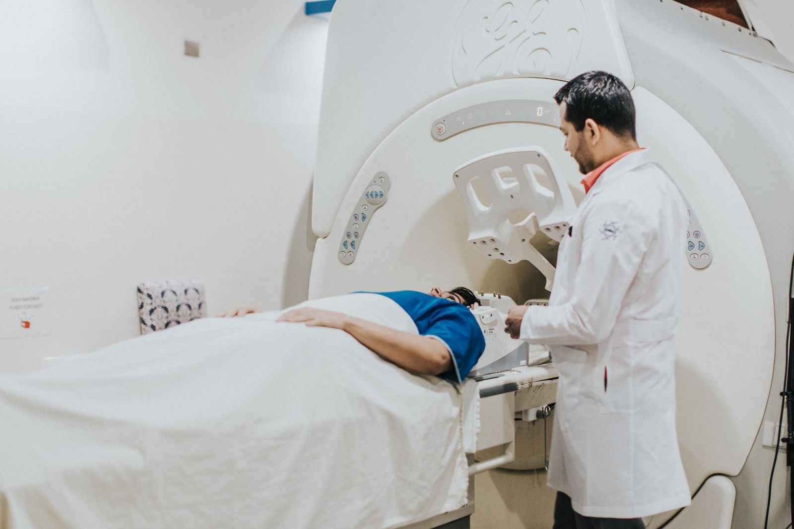
The NHS says an MRI scan usually takes between 15 minutes and 1 hour, and can take longer. The scanner makes loud bangs and clicks, so you will be offered ear plugs or headphones. You need to lie still, and before the scan you will be asked about any metal in your body, such as a pacemaker or surgical clips.
For more practical tips, see what to expect at an MRI scan and our guide to MRI options if you feel claustrophobic.
Where private MRI fits
Private MRI can have a role, but it is not a shortcut to an MS diagnosis. Some people who are already under a neurologist choose to have a requested scan privately. If you do, bring the request or referral letter so the clinic scans the right areas with the right technique, and make sure the report and images go back to your neurologist.
If you have had a private brain scan for another reason and the report mentions white matter changes, take it to your GP, who can decide whether a referral is needed. Our guide on getting your scan images and report explains how to share them.
Please speak to your GP, or the clinician who referred you, about any symptoms or results, and if symptoms are urgent, call NHS 111, or 999 in an emergency.
Finding a private scan
You can compare regulated clinics offering MRI on FindScan, see each clinic's own listed price, and send an enquiry to the clinic you choose. The clinic will contact you to confirm the details and the final price. MRI does not use ionising radiation, and some clinics accept self-referral while others ask for a referral from a doctor, so check with the clinic. If MS is suspected, your GP and neurologist should guide which scans you need.
Common questions
Can an MRI scan diagnose MS on its own?
No. NICE says MS should not be diagnosed on the basis of MRI findings alone. A neurologist combines your history, an examination, MRI and sometimes laboratory tests, following the McDonald criteria. MRI is very important, and the MS Society says it confirms the diagnosis in over 90 per cent of people with MS, but it is always interpreted alongside your symptoms.
Can an MRI rule out MS?
Not by itself. There is no single test for MS. Most people with MS have changes on MRI, but not everyone does. The MS Trust says that if scans do not show typical lesions, diagnosis is harder and a neurologist may repeat scans after a period of waiting. If you still have symptoms, keep talking to your GP or neurologist.
What do MS lesions look like on MRI?
On a common type of image called a T2 scan, MS lesions show up as bright spots where the myelin coating around nerves has been damaged. If contrast dye is used, active lesions may light up, showing current inflammation. Similar bright spots can also be caused by migraine, stroke or normal ageing, so a specialist needs to interpret them.
Will I need contrast dye for an MS MRI scan?
Not always. The MS Trust says some people are asked to have an extra scan with an injection of gadolinium dye, which helps tell new or growing lesions apart from older scarring. Your neurologist and the radiology team decide whether contrast is needed. The clinic will ask some safety questions before giving it.
Should I get a private MRI if I think I have MS?
Start with your GP. NICE recommends that anyone suspected of having MS is referred to a neurologist for diagnosis, and a scan alone cannot diagnose MS. If your neurologist requests an MRI and you choose to have it privately, take the request with you and make sure the report and images go back to them.
Why would I need follow-up MRI scans for MS?
Follow-up scans can show whether new or active lesions have appeared over time. The MS Trust says MRI is used to monitor relapses and progression, and lesions can appear on MRI before they cause symptoms. If the diagnosis is not yet clear, a repeat scan can also help show change over time. Your neurologist decides when scans are needed.
References
- Multiple sclerosis. NHS. Accessed 12 October 2026.
- Multiple sclerosis in adults: management (NG220), Recommendations. NICE. Accessed 12 October 2026.
- Magnetic resonance imaging (MRI). MS Trust. Accessed 12 October 2026.
- Lesion (MS lesion). MS Trust. Accessed 12 October 2026.
- How is MS diagnosed?. MS Trust. Accessed 12 October 2026.
- How is MS diagnosed? Tests for MS. MS Society. Accessed 12 October 2026.
- MRI scan. NHS. Accessed 12 October 2026.
- Diagnosis of MS. Norfolk and Norwich University Hospitals NHS Foundation Trust. Accessed 12 October 2026.
This article is general information, not medical advice, and it does not replace an assessment by a clinician. If you have symptoms that worry you, speak to your GP or call 111. In an emergency, call 999.