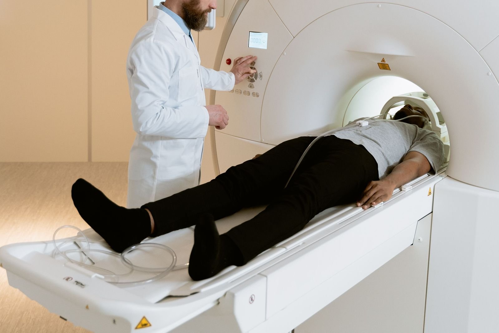
Chest MRI Scan: What an MRI of the Chest Looks At, How It Compares With CT and X-ray, and What to Expect
A chest MRI is a specialised scan, usually chosen by a specialist to answer a particular question about the heart, the large blood vessels, the chest wall or the middle of the chest. This guide explains what it shows, why the lungs are usually checked with CT instead, and what happens before, during and after the scan.
Written by the FindScan Editorial Team and reviewed by our Medical Advisor, Dr Mohamed Najjar
Reviewed
A chest MRI uses a strong magnet and radio waves, rather than X-rays, to take detailed pictures inside the chest. People search for it as "MRI of chest" or "MRI chest scan", and the NHS describes an MRI scan simply as a test that takes detailed pictures of the inside of your body. In the chest, MRI is very good at showing the heart, the large blood vessels, the chest wall and the area in the middle of the chest, and less suited to the lungs themselves, where CT is usually the main test.
This guide explains what a chest MRI looks at, the common reasons a specialist asks for one, how it compares with chest CT and chest X-ray, and what happens on the day. If you already have a referral or a clear clinical question, you can compare private MRI clinics on FindScan.
What a chest MRI looks at
The chest holds the heart, the aorta and the other large blood vessels, the lungs, the chest wall (ribs, muscles and soft tissue) and the mediastinum, which is the space in the middle of the chest between the two lungs. A review of chest MRI in the journal Radiologia Brasileira says MRI plays a role in assessing heart and blood vessel disease and lesions in the mediastinum, and in checking whether a tumour has grown into the mediastinum, the lining of the lungs (pleura) or the chest wall. It adds that clinical use is generally restricted to specific conditions.
When the question is mainly about the heart, the scan is usually set up as a cardiac MRI. The British Heart Foundation describes this as a scan of your heart and blood vessels that can help check for problems such as congenital heart disease, cardiomyopathy, valve disease and heart muscle damage.
A breast MRI is a separate test, requested for its own reasons, so a chest MRI is not a way to check the breasts. Our guide to breast MRI and mammograms explains when that scan is used.
Why an MRI chest scan is requested
The leaflet from University Hospital Southampton on cardiac and chest MRI says MRI can be used to help diagnose conditions, plan treatments and assess how well previous treatment has worked. It advises asking the doctor who referred you if you are not sure why you need the scan.
Reasons described in the chest MRI review include:
- looking more closely at a mass or lump in the mediastinum
- checking whether a known tumour has spread into the chest wall, pleura or mediastinum
- problems of the chest wall and of the nerves at the top of the chest that supply the arm (the brachial plexus)
- heart and blood vessel conditions, including some cases of high blood pressure in the lungs (pulmonary hypertension)
- follow-up scans where repeated radiation is a concern, such as in young people and in cystic fibrosis
The NHS page on diagnosing lung cancer lists a CT scan among the main tests, and says that once lung cancer has been diagnosed you may need more tests, which can include an MRI scan. In practice, a chest MRI is often a follow-on test chosen by a specialist after an X-ray or CT, rather than the first scan.
MRI of chest, chest CT or chest X-ray: how they compare
Chest X-ray. The NHS says an X-ray uses a small amount of radiation, is used mainly to check bones, teeth and lungs, and should only take a few minutes. It is often the first test for chest symptoms. See our chest X-ray page and our guide to what a chest X-ray can show.
Chest CT. CT gives detailed cross-section images and is the usual scan for the lungs. The chest MRI review explains why MRI struggles there: lung tissue gives off very little signal, the many tiny air pockets distort the image, and breathing and the heartbeat cause constant movement. Our chest CT scan page and CT chest guide explain that test.
Chest MRI. MRI does not use ionising radiation and shows soft tissue in fine detail, which is why it suits the heart, blood vessels, chest wall and mediastinum. The same review notes it is valuable where radiation is a particular concern, such as in young patients. Our guide to MRI vs CT compares the two in more depth.
X-ray and CT both use ionising radiation. The Care Quality Commission says the IR(ME)R regulations apply in both the NHS and the independent sector, name the referrer as one of the duty holders, and require each exposure to be justified so the benefits outweigh the risks. So a chest X-ray or chest CT always needs a referrer; many private clinics can arrange this through their own doctor.
Preparing for a chest MRI: safety screening and what to wear
Because MRI uses a very strong magnet, every clinic checks your safety first. The NHS says you will fill in a form asking whether you are pregnant or breastfeeding, have had surgery, have kidney problems, have metal parts in your body such as surgical clips, a pacemaker, dental fillings or bridges, or have any allergies, including to contrast dye. It says not to wear anything with metal in it, such as jewellery, a bra, hair clips or hearing aids, and that you may change into a hospital gown.
The Southampton chest MRI leaflet says you can eat and drink normally before the scan, and asks you to remove all jewellery, piercings, drug patches, metallic denture plates and hearing aids. It asks you to contact the team beforehand if you have a pacemaker or defibrillator, a heart valve replacement, metal in your eyes, any metallic or electronic implant, surgery in the past six weeks, or have swallowed a capsule camera, and warns that the scan may have to be postponed if you do not.
Royal Papworth Hospital says many people with artificial joints or coronary stents can be scanned, but the team needs to know beforehand so the device can be checked, and suggests avoiding sportswear because its fibres can affect the scan. Our guides to MRI safety with metal implants and claustrophobia and MRI cover common worries.
Contrast, breath-holds and how long a chest MRI takes
Some chest scans use contrast dye. The Southampton leaflet says a thin plastic tube (cannula) may be placed in a vein in your arm if needed, and that contrast helps certain tissues and blood vessels show up more clearly. Royal Papworth asks you to tell the team beforehand if you have kidney problems. The NHS says side effects of contrast are usually mild and should not last long. Our guide to MRI with contrast explains more.
Breathing moves the chest, so you will be asked to hold your breath for a few seconds at a time, with instructions given through headphones. For scans that include the heart, small sticky pads are placed on your chest so the scanner can take pictures in time with your heartbeat.
Southampton says a chest MRI takes approximately 30 to 40 minutes and a cardiac MRI approximately one hour. The NHS says MRI scans usually take between 15 minutes and 1 hour but can take longer.
Results and referral for a chest MRI
A radiologist, a doctor who specialises in reading scans, looks at the images and writes a report for the doctor who referred you; our guide to radiographers and radiologists explains who does what. The NHS says it usually takes 1 to 2 weeks to get MRI results, and that if you have not heard anything after a few weeks you should contact the doctor who referred you. Our guide to how long scan results take covers the private route. A report that finds nothing abnormal answers the question that was asked; it does not rule out every condition, so go through it with your clinician.
MRI does not use ionising radiation, so some private clinics accept self-referral for some MRI scans, depending on the clinic's own policy and its safety screening. A chest MRI is a specialised test, though, and a clinic may ask for a referral letter or the clinical question before it agrees the scan, so it is protocolled correctly. Our guide to private MRI without a GP referral explains how this works.
When chest symptoms need urgent help
A scan is not the right first step for sudden chest symptoms. The NHS says to call 999 if you get sudden pain or discomfort in your chest that does not go away, pain that spreads to your arms, neck, jaw, stomach or back, or chest pain with sweating, feeling sick, light-headed or short of breath.
Comparing clinics on FindScan
On FindScan you can compare MRI clinics near you, see each clinic's own listed price and send an enquiry; the clinic then contacts you to arrange your scan and confirm whether it needs a referral. Not every MRI clinic offers chest or cardiac MRI, so check this with the clinic when you enquire.
FindScan is a directory of independent, regulated imaging clinics, not a healthcare provider, and nothing here is medical advice. Speak to your GP or the clinician who referred you about symptoms or results. If you need help urgently, call NHS 111, or 999 in an emergency.
Common questions
What does a chest MRI show?
A chest MRI shows soft tissue in detail, so it is mainly used for the heart, the large blood vessels, the chest wall and the mediastinum, the space between the lungs. A published review says it helps assess heart and blood vessel disease, masses in the mediastinum and whether a tumour has spread into the chest wall or lining of the lungs. The lungs themselves are usually checked with CT.
How long does an MRI chest scan take?
University Hospital Southampton says a chest MRI takes approximately 30 to 40 minutes, and a cardiac MRI approximately one hour. The NHS says MRI scans usually take between 15 minutes and 1 hour but can take longer. Allow extra time for the safety checks and changing, and for a cannula if contrast is needed.
MRI of chest or CT: which is better for the lungs?
For most lung questions, CT is the usual scan. Lung tissue gives very little MRI signal, the air pockets distort the image, and breathing and the heartbeat cause movement. MRI is better suited to the heart, blood vessels, chest wall and mediastinum, and avoids ionising radiation. The choice is for the specialist who knows the question being asked.
Do you need contrast for a chest MRI?
Not always. Some chest scans use a contrast dye given through a small tube in a vein in your arm, which helps certain tissues and blood vessels show up more clearly. The NHS says side effects are usually mild. Tell the clinic beforehand about any kidney problems or any allergy to contrast dye.
Is a chest MRI the same as a breast MRI?
No. A breast MRI is a separate test that is requested for its own reasons, so a chest MRI is not a way to check the breasts. A chest MRI looks at structures such as the heart, the large blood vessels, the chest wall and the middle of the chest. If you have a breast concern, speak to your GP first.
Can I refer myself for a chest MRI?
Some private clinics accept self-referral for some MRI scans, because MRI does not use ionising radiation, but this depends on the clinic's own policy and its safety screening. A chest MRI is specialised, so a clinic may ask for a referral letter or the clinical question first. Chest X-ray and chest CT always need a referrer, which many private clinics can arrange.
References
- MRI scan. NHS. Accessed 29 October 2026.
- Magnetic resonance imaging (MRI) cardiac and chest scan (patient information factsheet). University Hospital Southampton NHS Foundation Trust. Accessed 29 October 2026.
- Magnetic resonance imaging (MRI): patient information sheet (PI 5). Royal Papworth Hospital NHS Foundation Trust. Accessed 29 October 2026.
- MRI scan (cardiac MRI). British Heart Foundation. Accessed 29 October 2026.
- Lung cancer: diagnosis. NHS. Accessed 29 October 2026.
- X-ray. NHS. Accessed 29 October 2026.
- Chest pain. NHS. Accessed 29 October 2026.
- Souza CA. MRI of the chest: review of imaging strategies. Radiol Bras. 2015;48(6):V-VI. Radiologia Brasileira (peer-reviewed, PubMed Central). Accessed 29 October 2026.
- Ionising Radiation (Medical Exposure) Regulations (IR(ME)R). Care Quality Commission. Accessed 29 October 2026.
This article is general information, not medical advice, and it does not replace an assessment by a clinician. If you have symptoms that worry you, speak to your GP or call 111. In an emergency, call 999.