Pituitary MRI: Why It Is Not Just a Brain Scan, and When You Might Need One
The pituitary is a small gland at the base of the brain, and scanning it well takes a dedicated MRI protocol rather than a routine head scan. Here is what the scan involves, who is usually sent for one and why the hormone tests normally come first.
Written by the FindScan Editorial Team and reviewed by our Medical Advisor, Dr Mohamed Najjar
Reviewed
Each October the Pituitary Foundation runs Pituitary Awareness Month, and it says this year's focus is symptoms: the slow, easy-to-miss changes that can take years to be linked to a gland most people have never thought about. When a doctor does start to suspect the pituitary, an MRI of the gland is usually part of the work-up.
A pituitary MRI is not the same as a standard brain MRI. It is a focused scan of a small area, often with a contrast injection, normally requested by a specialist after hormone tests have pointed to a problem. This guide explains what it looks at, why the order of tests matters, what happens on the day and what to ask if you arrange one privately.
What the pituitary gland does
University Hospitals Coventry and Warwickshire describes the pituitary as a small, oval gland at the base of the brain, below the optic nerves, that releases hormones controlling other glands and body functions, including growth, milk production, thyroid function, how concentrated your urine is and childbirth. Because it controls so much, a change in the gland can show up almost anywhere in the body.
The same leaflet says most pituitary tumours are non-cancerous (benign) growths called adenomas, which are usually slow growing.
Why a pituitary MRI is a different scan
Our guide to what a brain MRI shows lists a dedicated pituitary protocol among the extra sequences added when a scan has a particular question to answer. A routine brain MRI covers the whole head; a pituitary study concentrates on one small area. The Royal Berkshire NHS Foundation Trust's acromegaly leaflet describes it as a special MRI scan to look at the exact size of your pituitary gland, adding that you may be given an injection before the scan to improve the results.
University Hospitals Coventry and Warwickshire puts it simply: the scan will show the exact position of the tumour. That precision matters because the gland sits close to the optic nerves.
Who is usually sent for one
The clearest examples on NHS pages are hormone conditions. The NHS says acromegaly is usually caused by an adenoma in the pituitary gland, that symptoms such as swollen hands and feet and headaches develop slowly, and that a GP who suspects it may refer you to a specialist for tests including an MRI scan of your brain to see if you have a tumour. For Cushing's syndrome, the NHS says a small, non-cancerous growth in the pituitary gland is one cause, and that specialist tests may include scans of your brain and kidneys.
Prolactin-releasing tumours are the most common type, according to the Coventry and Warwickshire leaflet, which says they can stop monthly periods, cause breast milk production and affect fertility. The Royal Liverpool and Broadgreen Hospitals prolactinoma leaflet says that initially your prolactin level is checked and an MRI scan of your pituitary gland is performed. The Coventry leaflet also lists vision problems and headaches from pressure on the optic nerve, and the NHS brain tumours page says to see a GP if you have symptoms such as a headache that feels different from your usual headaches or headaches that are getting worse.
Why the hormone tests come first
In every NHS description above, the order is the same: hormone tests first, then a scan if they point to the pituitary. The Coventry and Warwickshire leaflet says the hormone tests come first and that, if levels are abnormal, further tests may include a CT or MRI brain scan and eye visual field tests. For acromegaly, the Royal Berkshire leaflet describes a glucose tolerance test over a period of about two hours as well as the MRI.
There is a practical reason for the order. A scan cannot say whether a gland is overproducing or underproducing a hormone. The hormone results tell the specialist what question the scan needs to answer, which decides the sequences and whether contrast is used. Our guide to seeing a GP before a scan covers the general principle.
What happens on the day, and afterwards
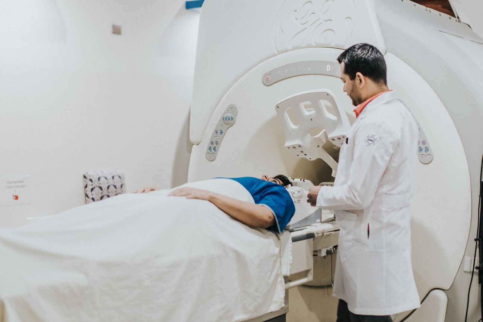
The scan itself is an MRI like any other. The NHS says an MRI usually takes between 15 minutes and 1 hour but can take longer, that you will need to lie still, and that beforehand you will fill in a form about things such as pregnancy, kidney problems, metal in your body such as a pacemaker, and allergies including to contrast dye. The Coventry and Warwickshire leaflet is blunter: if you have a pacemaker you cannot have an MRI scan, and any other metal implant should be discussed with the doctor well before the test. MRI departments work to safety guidance such as the MHRA's safety guidelines for MRI equipment, updated on 30 September 2026.
If contrast is used, the NHS says you may be asked to stay for up to 30 minutes afterwards to check you do not have a reaction; see MRI with contrast. Your head rests in a coil for a pituitary scan, which some people find more enclosed than a scan of a knee. The NHS says to speak to staff beforehand if you feel anxious or have claustrophobia, and that you may be offered medicine to help you relax; see claustrophobia and MRI.
For many pituitary conditions the first scan is not the last. The Royal Liverpool leaflet says the tablets used for prolactinomas reduce prolactin output and over time can also reduce the size of the tumour, that MRI or CT scans are repeated at regular intervals to check whether it is shrinking, and that discharge from clinic is considered when prolactin is normal off medication and the MRI shows the tumour has shrunk.
Arranging a pituitary MRI privately
MRI does not use ionising radiation, so many private clinics accept self-referral after their own safety screening. For the pituitary, though, the useful route is a referral from the endocrinologist or GP managing your hormone results, because the clinic needs to know the question, whether contrast is wanted and who should receive the report. When you enquire, ask whether the clinic offers a dedicated pituitary protocol, whether contrast is included, who reports the scan and how the report reaches your specialist.
You can compare brain and head MRI clinics near you and send an enquiry; the clinic contacts you to confirm the referral it needs and the next steps. FindScan is a directory of independent, regulated imaging clinics: you compare clinics and send an enquiry, and the clinic contacts you. We are not a healthcare provider, and nothing here replaces advice from your GP or specialist.
Some symptoms need urgent help, not a scan. The NHS says to call 999 or go to A&E if a headache started suddenly and is extremely painful, or comes with loss of vision, numbness or weakness, difficulty speaking, drowsiness or confusion, or a seizure.
Common questions
Is a pituitary MRI the same as a brain MRI?
No. A brain MRI covers the whole head. A pituitary MRI concentrates on the small gland at the base of the brain and, according to the Royal Berkshire NHS Foundation Trust, may include an injection to improve the results.
Do I need contrast for a pituitary MRI?
Often, yes. NHS trust leaflets describe an injection before the scan to improve the pictures, and the NHS says you may be asked to stay up to 30 minutes afterwards to check for a reaction. Whether contrast is used depends on the question being asked.
Can I have a pituitary MRI without a referral?
Many private clinics accept self-referral for MRI, but a pituitary scan is almost always requested after hormone tests by an endocrinologist or GP, who decides the protocol and reads the report alongside those results.
Does the scan show my hormone levels?
No. The MRI shows the size and position of the gland and any growth in it. Hormone levels come from tests arranged by your doctor, which the Coventry and Warwickshire leaflet says come first.
A scan I had for another reason mentioned my pituitary. What should I do?
Take the report to the doctor who requested the scan, or to your GP, who can decide whether hormone tests or an endocrinology referral are needed. A mention on a report is a reason for a conversation, not a reason to arrange another scan yourself.
References
- October is Pituitary Awareness Month. The Pituitary Foundation. Accessed 27 October 2026.
- Pituitary tumours (patient information leaflet). University Hospitals Coventry and Warwickshire NHS Trust. Accessed 27 October 2026.
- Acromegaly. NHS. Accessed 27 October 2026.
- Cushing's syndrome. NHS. Accessed 27 October 2026.
- Prolactinoma (patient information leaflet). Royal Liverpool and Broadgreen University Hospitals NHS Trust. Accessed 27 October 2026.
- Acromegaly (patient information leaflet, January 2025). Royal Berkshire NHS Foundation Trust. Accessed 27 October 2026.
- Brain tumours. NHS. Accessed 27 October 2026.
- MRI scan. NHS. Accessed 27 October 2026.
- Headaches. NHS. Accessed 27 October 2026.
- Magnetic resonance imaging equipment in clinical use: safety guidelines. MHRA (GOV.UK). Accessed 27 October 2026.
This article is general information, not medical advice, and it does not replace an assessment by a clinician. If you have symptoms that worry you, speak to your GP or call 111. In an emergency, call 999.