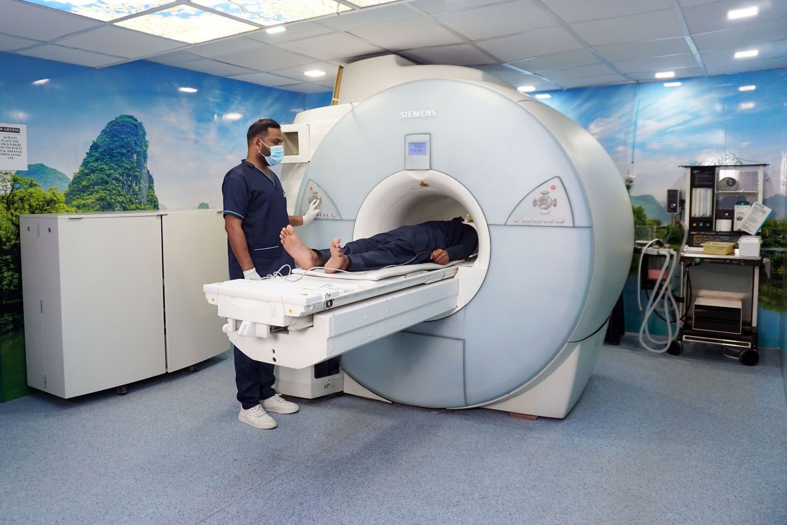
MRA Scan (MR Angiogram): What It Shows, MRA vs MRI, and Having a Private MRA Scan
An MRA scan is an MRI scan of your blood vessels. This guide explains what an MR angiogram is used for, how it compares with a standard MRI, a CT angiogram, a catheter angiogram and ultrasound, and what to know before a private MRA scan.
Written by the FindScan Editorial Team and reviewed by our Medical Advisor, Dr Mohamed Najjar
Reviewed
An MRA scan, short for magnetic resonance angiogram, is a type of MRI scan that looks at your blood vessels and the blood flowing through them. The Princess Alexandra Hospital NHS Trust patient leaflet says it is used to find diseased vessels and vessel malformations such as aneurysms, to help plan surgery and to monitor existing conditions. If you are comparing MRA vs MRI, or thinking about a private MRA scan, the key point is that an MR angiogram is a specific test that answers a specific question about blood vessels.
This guide covers what an MRA shows, when contrast dye is used, how it compares with other ways of looking at blood vessels, the safety checks, how long it takes and how results reach you. It also explains when symptoms need 999 rather than a scan, and how referral works for private MRI scans.
What is an MRA scan, and is contrast used?
An MRA uses the same kind of scanner as any other MRI, so it does not use ionising radiation. The NHS page on angiography explains that angiography is traditionally a type of X-ray used to check blood vessels, but that it can be done using scans instead, called CT angiography or MRI angiography. An MRA is the MRI version.
The PAH leaflet says the vessels most often examined with MRA are those in the head and neck, the kidneys, the legs and the pelvis.
Some MRA scans need contrast and some do not. The leaflet says an MRI sometimes requires an injection of a special dye, given through a vein in the arm or hand when the radiologist asks for it, and that the dye is cleared from the body by the kidneys. It says allergic reactions are possible but rare, and that the dye can occasionally leak into the tissue around the injection site and cause swelling. Breastfeeding can continue as normal afterwards.
The dye used for contrast-enhanced MRA is gadolinium-based. AHealth Research Authority summary of an NHS study says most MRI centres use contrast-enhanced MRA as their tried and tested technique, while non-contrast alternatives can also give reliable, high quality images. In that study the contrast technique still scored best for the arteries to the kidneys, so the choice depends on the question being asked. Our guide to MRI with contrast explains the dye in more detail.
What an MR angiogram is used for
Brain aneurysm follow-up. The NHS describes a brain aneurysm as a swelling in a blood vessel in the brain. It says most are small and cause no symptoms, and that they are often found during a test for another condition. Treatment is not usually needed for small aneurysms that are not causing symptoms, and you may just need regular follow-up scans to check whether it is getting bigger.
For people who have had a bleed from an aneurysm, NICE guideline NG228 says the choice of follow-up scan, and how often and for how long it is done, should be based on things like the treatment already given, the estimated risk of further bleeding and the person's own preference. The type of follow-up scan is a decision for your specialist team.
Neck (carotid) arteries. After a transient ischaemic attack (TIA), the NHS says a carotid ultrasound scan can show if there is narrowing or a blockage in the neck arteries leading to the brain. MRA of the head and neck is another way of imaging these vessels, and your specialist will choose the test.
Leg (peripheral) arteries. For peripheral arterial disease, NICE guideline CG147 recommends duplex ultrasound as the first test when treatment to restore blood flow is being considered. It then recommends contrast-enhanced MRA for people who need further imaging, and CT angiography if MRA is not suitable or not tolerated.
Kidney arteries. The PAH leaflet lists the kidney vessels among those commonly examined. If your doctor has suggested a kidney MRA, ask them what question the scan is meant to answer.
MRA vs MRI, CT angiogram, catheter angiogram and ultrasound
MRA vs MRI. Both use the same scanner. A standard brain MRI is set up to show brain tissue, while an MRA is set up to show the blood vessels. Your referral should say which one is needed, or whether both are, because the clinic plans the scan around it.
CT angiogram. This uses a CT scanner and a contrast injection. The NHS CT scan page says a CT scan usually takes 10 to 20 minutes, and that if you have lots of CT scans there is a very small chance the radiation can cause cancer, which is why doctors weigh up the risks first. NICE says CT angiography of the head should be offered without delay to people with a confirmed bleed on the brain. Because CT uses ionising radiation, it always needs a referrer. Our guide to the CT coronary angiogram covers the heart version.
Catheter angiogram. The NHS says a very thin flexible tube is put into an artery and guided to the area being checked, and X-ray pictures are taken. It is generally safe and painless, but there is a very small risk of more serious complications, such as an allergic reaction, stroke or heart attack. It can take 30 minutes to 2 hours. NICE says it can be considered if a CT angiogram has not found the cause of a bleed, with MRA used instead if a catheter angiogram is not suitable.
Ultrasound. Ultrasound uses sound waves and no radiation. It is the test the NHS describes for checking the neck arteries after a TIA, and the first-line test NICE recommends for leg arteries. Our guide to the carotid ultrasound scan explains what it involves.
Urgent symptoms: call 999, do not wait for a scan
If you think someone is having a stroke, use the word FAST from the NHS stroke page: face, arms, speech and time to call 999. The NHS says to call 999 right away, including if stroke signs happened in the last 24 hours and have stopped. Do not drive yourself to A&E.
The NHS brain aneurysm page says to call 999 if you or someone else suddenly gets an extremely painful headache that does not go away, weakness in the face, arm or body, numbness, speech problems, or blurred vision or loss of sight. NICE describes a thunderclap headache as a sudden severe headache that typically peaks within 1 to 5 minutes. These are emergencies, not reasons to look for a private scan.
If you keep getting headaches, or they are not going away or are getting worse, the NHS says to ask for an urgent GP appointment or contact NHS 111.
Safety screening, how long an MRA scan takes, and results
Before any MRI you fill in a safety form. The PAH leaflet says that if you have a pacemaker or another programmable implant, that trust cannot scan you, so always tell the clinic about any implant before the day. It also asks you to get in touch about jewellery or piercings you cannot remove, metal fragments such as shrapnel, and fixed braces. Fillings and crowns are fine. If you are or may be pregnant, it says the decision to scan is made with your referring clinician and the radiologist. Our guide to MRI safety and metal implants has more.
The leaflet says the scan is painless and typically takes 15 to 60 minutes. You lie still on the table, wear ear protection because the scanner is loud, and hold a call button so you can stop the scan if you need to.
A radiologist reports the scan to the clinician who arranged it, who then discusses the results with you. The leaflet says this usually takes a couple of weeks, and that you may occasionally be asked back for more images. A report that does not find a problem answers the question the scan was asked; it is not a general health check. See our guide on how long scan results take.
Do you need a referral for a private MRA scan?
MRI does not use ionising radiation, so some clinics accept self-referral for MRI, depending on the clinic's own policy and its safety screening. Others ask for a referral from a GP or specialist. Our guide to private MRI without a GP referral explains how this works.
Scanning people without symptoms for aneurysms is a different matter. NICE says routine testing for aneurysms in the relatives of people who have had a bleed has not been shown to save lives or prevent bleeds. It says testing should be based on the relative's own risk and is usually limited to people with at least two first-degree relatives (a parent, brother or sister) who have had a bleed from an aneurysm. Whether an MRA is worthwhile without symptoms is a decision to make with a clinician. Our guide to the limits of health scans explains why.
If you already have a known aneurysm under follow-up, agree any private scan with your specialist team first, so the images and report can be used by the people looking after you.
Finding a private scan
Each clinic on FindScan lists its own price and confirms the final price when it contacts you. When you send an enquiry, say that you need an MRA, which area is being checked and whether you have a referral letter. MRI may be self-referral at some clinics, depending on the clinic, while a CT angiogram always needs a referrer.
Comparing clinics on FindScan
On FindScan you can compare MRI clinics near you, see what each clinic offers, and send an enquiry to the one you choose. The clinic contacts you to confirm whether it performs MRA, whether it needs a referral, and the next steps to arrange your scan.
FindScan is a directory of independent, regulated imaging clinics. We are not a healthcare provider and nothing here is medical advice, so please speak to your GP or the clinician who referred you about your symptoms or results. If symptoms are urgent, call NHS 111, or 999 in an emergency.
Common questions
What is an MRA scan?
An MRA scan, or magnetic resonance angiogram, is an MRI scan that looks at your blood vessels and blood flow. NHS patient information says it is used to find diseased vessels and malformations such as aneurysms, to help plan surgery and to monitor existing conditions. The vessels most often checked are in the head and neck, kidneys, legs and pelvis.
MRA vs MRI: what is the difference?
Both use the same MRI scanner and neither uses ionising radiation. A standard MRI is set up to show organs and tissue, such as the brain. An MRA is set up to show the blood vessels. A referral may ask for one or both, so check with the clinic what your referral says before your appointment.
Is an MR angiogram done with contrast?
Sometimes. NHS patient information says an MRI sometimes needs an injection of a special dye through a vein in the arm or hand, which the kidneys then clear from the body. Allergic reactions are possible but rare. Non-contrast MRA techniques also exist, and the radiologist chooses the approach that best answers the question being asked.
Can I get a private MRA scan without a GP referral?
MRI does not use ionising radiation, so some clinics accept self-referral, depending on the clinic's own policy and safety screening. Others ask for a GP or specialist referral. NICE says routine testing for aneurysms in relatives of people who had a bleed has not been shown to save lives, so scanning without symptoms is a decision to make with a clinician.
How long does an MRA scan take?
NHS patient information says an MRI angiogram typically takes 15 to 60 minutes. You need to lie still, you wear ear protection because the scanner is loud, and you hold a call button so you can stop the scan. A radiologist reports the scan, and results usually reach the clinician who arranged it within a couple of weeks.
MRA vs CT angiogram: which is better?
Neither is better in every case. A CT angiogram is quick and is what NICE recommends without delay for a confirmed bleed on the brain, but it uses ionising radiation and always needs a referrer. MRA uses no radiation. For leg arteries, NICE recommends ultrasound first, then contrast-enhanced MRA, with CT angiography if MRA is not suitable.
References
- MRI Angiogram scan / Angiography: patient information leaflet (reviewed February 2026). The Princess Alexandra Hospital NHS Trust. Accessed 29 October 2026.
- Angiography. NHS. Accessed 29 October 2026.
- Brain aneurysm (reviewed 9 October 2025). NHS. Accessed 29 October 2026.
- Subarachnoid haemorrhage caused by a ruptured aneurysm: diagnosis and management (NG228), recommendations. NICE. Accessed 29 October 2026.
- Peripheral arterial disease: diagnosis and management (CG147), guidance. NICE. Accessed 29 October 2026.
- Stroke: symptoms (reviewed 12 September 2024). NHS. Accessed 29 October 2026.
- Transient ischaemic attack (TIA): diagnosis. NHS. Accessed 29 October 2026.
- CT scan. NHS. Accessed 29 October 2026.
- Reducing Gadolinium Based Contrast Agents in MRA (research summary). Health Research Authority. Accessed 29 October 2026.
This article is general information, not medical advice, and it does not replace an assessment by a clinician. If you have symptoms that worry you, speak to your GP or call 111. In an emergency, call 999.